 Explore
 Validate
 Learn
Learn Western blot
 Immunohistochemistry
Antibody data
- Antibody Data
- Antigen structure
- References [3]
- Comments [0]
- Validations
- Immunohistochemistry [1]
- Flow cytometry [1]
- Other assay [4]
Submit
Validation data
Reference
Comment
Report error
- Product number
- PA5-23124 - Provider product page

- Provider
- Invitrogen Antibodies
- Product name
- TLR4 Polyclonal Antibody
- Antibody type
- Polyclonal
- Antigen
- Synthetic peptide
- Description
- Suggested positive control: recombinant protein, RAW, Daudi.
- Reactivity
- Human, Mouse, Rat
- Host
- Rabbit
- Isotype
- IgG
- Vial size
- 100 µg
- Concentration
- 1 mg/mL
- Storage
- Store at 4°C short term. For long term storage, store at -20°C, avoiding freeze/thaw cycles.
Submitted references Deficiency of inhibitory TLR4 homolog RP105 exacerbates fibrosis.
LPS alters the immuno-phenotype of glioma and glioma stem-like cells and induces in vivo antitumor immunity via TLR4.
Association between toll-like receptor 4 expression and neural stem cell proliferation in the hippocampus following traumatic brain injury in mice.
Wang W, Bale S, Yalavarthi B, Verma P, Tsou PS, Calderone KM, Bhattacharyya D, Fisher GJ, Varga J, Bhattacharyya S
JCI insight 2022 Nov 8;7(21)
JCI insight 2022 Nov 8;7(21)
LPS alters the immuno-phenotype of glioma and glioma stem-like cells and induces in vivo antitumor immunity via TLR4.
Han S, Wang C, Qin X, Xia J, Wu A
Journal of experimental & clinical cancer research : CR 2017 Jun 22;36(1):83
Journal of experimental & clinical cancer research : CR 2017 Jun 22;36(1):83
Association between toll-like receptor 4 expression and neural stem cell proliferation in the hippocampus following traumatic brain injury in mice.
Ye Y, Xu H, Zhang X, Li Z, Jia Y, He X, Huang JH
International journal of molecular sciences 2014 Jul 17;15(7):12651-64
International journal of molecular sciences 2014 Jul 17;15(7):12651-64
No comments: Submit comment
Supportive validation
- Submitted by
- Invitrogen Antibodies (provider)
- Main image

- Experimental details
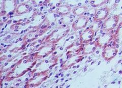
- Immunohistochemical analysis of TLR4 in mouse kidney tissue using a TLR4/CD284 polyclonal antibody (Product # PA5-23124) at 5 µg/mL.
Supportive validation
- Submitted by
- Invitrogen Antibodies (provider)
- Main image
- Experimental details
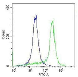
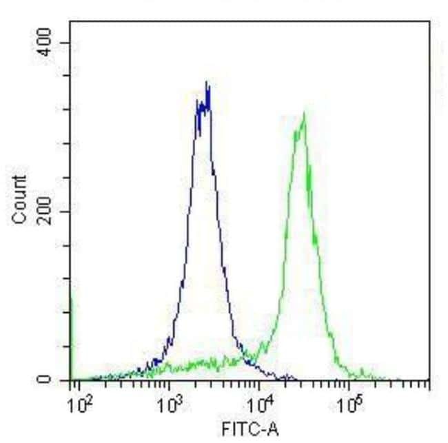
- Flow Cytometry analysis of TLR4 in formaldehyde-fixed THP-1 cells (green) compared to an isotype control (blue). Staining was performed using a TLR4 polyclonal antibody (Product # PA5-23124) at a dilution of 20µg x 10^6 cells., followed by secondary detection using a Goat anti-Rabbit IgG (H+L) Cross Adsorbed Secondary Antibody, DyLight 488 conjugate (Product # A-11008) at a dilution of 1:200. Rabbit IgG Isotype Control (Product # MA5-16385) was used as a negative control at the same dilution as the primary antibody. Data provided courtesy of Antibody Resource.
Supportive validation
- Submitted by
- Invitrogen Antibodies (provider)
- Main image

- Experimental details
- NULL
- Submitted by
- Invitrogen Antibodies (provider)
- Main image

- Experimental details
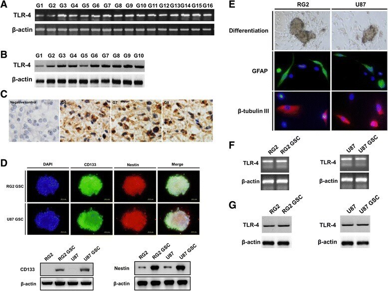
- Fig. 1 TLR4 expression in glioma cells. Representative images are shown. a - b : TLR4 mRNA and protein is expressed in glioblastoma clinical samples as determined by RT-PCR ( a ) and western blot ( b ). c : TLR4 protein expression and localization was examined using immunohistochemistry in glioblastoma. d : Neurospheres comprised of CD133 and nestin-positive glioma stem-like cells (GSCs) isolated from RG2 and U87 cells. The expression of CD133 and nestin was examined and compared in GSCs and the parental cells by western blot. e : GSCs became adherent and differentiated into GFAP- or beta III tubulin-positive cells. f - g : The expression of TLR4 in RG2 and U87 GSCs was determined by RT-PCR ( f ) and western blot ( g )
- Submitted by
- Invitrogen Antibodies (provider)
- Main image
- Experimental details
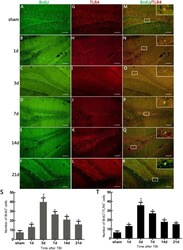
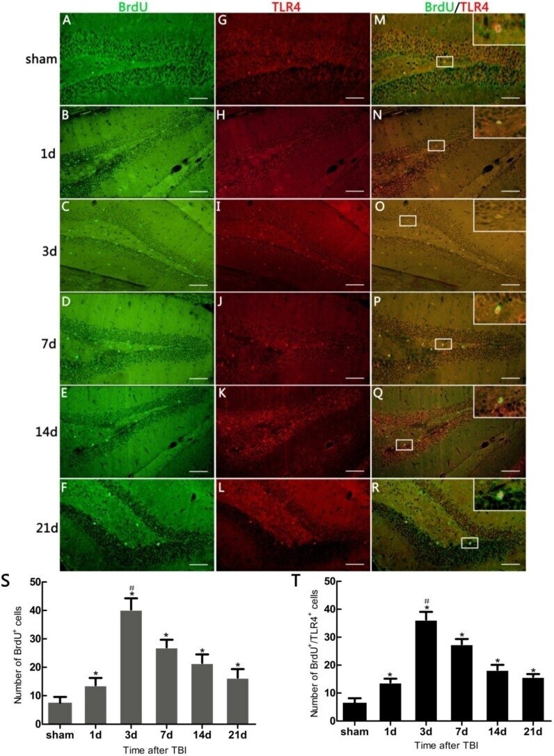
- Figure 2 Immunofluorescence (IF) microphotographs in dentate gyrus (DG) of the hippocampus in sham- and TBI-mice with various survivals post-injury. ( A - F ) 5-bromo-2-deoxyuridine (BrdU) (green) indicating neural stem cells (NSCs) in DG; ( G - L ) Toll-like Receptor 4 (TLR4) (red) indicating TLR4 expression in DG; ( M - R ) Merged IF of BrdU/TLR4 indicating TLR4 expression on the proliferating cells (suggestive of NSC proliferation), the rectangle area, taken from a magnified optic field, showing BrdU/TLR4 expressed in/on a newly-generated cell; ( S )The number of BrdU + cells revealing the degree to which putative NSCs have proliferated in DG varies over different survivals post-injury. TBI-mice has significantly more BrdU + cells than the sham ( * p < 0.05), and shows an obvious difference in the number of these cells between all post-traumatic time-points ( # p < 0.05); ( T ) The number of BrdU + /TLR4 + cells revealing the degree of TLR4 expression in the proliferating cells in DG varies over different survivals post-injury. TBI-mice has significantly more BrdU + /TLR4 + cells than the sham ( * p < 0.05), and shows an obvious difference in the number of these cells between all posttraumatic time-points ( # p < 0.05). Scale bar: 50 mum. Data are presented as Mean +- SD, n = 6. SD: standard deviation, d: day.
- Submitted by
- Invitrogen Antibodies (provider)
- Main image
- Experimental details
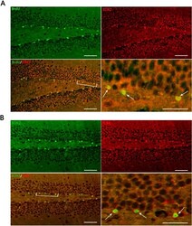
- Figure 3 Representative IF microphotographs in the DG of hippocampus in sham- and TBI-mice with various post-injury survival periods. ( A ) BrdU (green), indicating proliferating cells in DG, SOX2 (red), indicating NSCs in DG; merged IF of BrdU/SOX2 indicates that the proliferating cells are presumed, morphologically, to be NSCs (the percentage of putative NSCs in proliferating cells was 95.80% +- 7.91%); the arrows indicate BrdU + /SOX2 + cells in DG; ( B ) SOX2 (green) indicating NSCs in DG; TRL4 (red), indicating TLR4 expression in DG; merged IF of SOX2/TLR4, indicates TLR4 expression on NSCs, the arrows indicating SOX2 + /TLR4 + cells in DG. The bracketed area is magnified respectively as the right lower part in ( A ) and ( B ). Scale bar: 50 mum; Data are presented as Mean +- SD, n = 6.
