Explore
 Validate
 Learn
Learn Western blot
 Immunohistochemistry
 Flow cytometry
Antibody data
- Antibody Data
- Antigen structure
- References [1]
- Comments [0]
- Validations
- Flow cytometry [5]
- Other assay [2]
Submit
Validation data
Reference
Comment
Report error
- Product number
- PA5-23125 - Provider product page

- Provider
- Invitrogen Antibodies
- Product name
- TLR4 Polyclonal Antibody
- Antibody type
- Polyclonal
- Antigen
- Synthetic peptide
- Reactivity
- Human, Mouse, Porcine
- Host
- Rabbit
- Isotype
- IgG
- Vial size
- 100 μg
- Concentration
- 1 mg/mL
- Storage
- -20°C, Avoid Freeze/Thaw Cycles
Submitted references Electroacupuncture Improved Hippocampal Neurogenesis following Traumatic Brain Injury in Mice through Inhibition of TLR4 Signaling Pathway.
Ye Y, Yang Y, Chen C, Li Z, Jia Y, Su X, Wang C, He X
Stem cells international 2017;2017:5841814
Stem cells international 2017;2017:5841814
No comments: Submit comment
Supportive validation
- Submitted by
- Invitrogen Antibodies (provider)
- Main image
- Experimental details
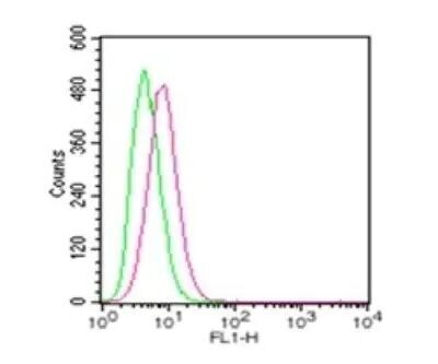
- Cell surface flow cytometric analysis using TLR4/CD284 polyclonal antibody (Product # PA5-23125). Mouse macrophage cells were probed using 1 µg of TLR4/CD284 polyclonal antibody (Product # PA5-23125) (red) and 1 µg of isotype control antibody (green). PE-conjugated secondary were used for this test.

- Submitted by
- Invitrogen Antibodies (provider)
- Main image

- Experimental details
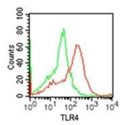
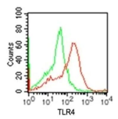
- Flow cytometry of TLR4 in RAW cells. Samples were incubated in TLR4 polyclonal antibody (Product # PA5-23125) using a dilution of 2 µg. Antibody (conjugated to Alexa Fluor ® 488) (red) and 2 µg of isotype control antibody (green).

- Submitted by
- Invitrogen Antibodies (provider)
- Main image

- Experimental details
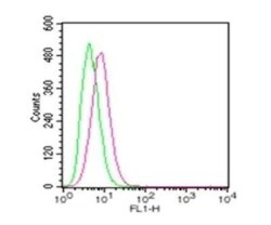
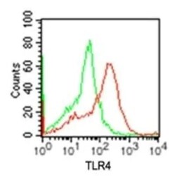
- Flow cytometry of TLR4 in Mouse macrophage cells. Samples were incubated in TLR4 polyclonal antibody (Product # PA5-23125) using a dilution of 1 µg followed by PE-conjugated secondary. Antibody (red) and 1 µg of isotype control antibody (green).

- Submitted by
- Invitrogen Antibodies (provider)
- Main image
- Experimental details
- Flow cytometry of TLR4 in RAW cells. Samples were incubated in TLR4 polyclonal antibody (Product # PA5-23125) using a dilution of 2 µg. Antibody (conjugated to Alexa Fluor ® 488) (red) and 2 µg of isotype control antibody (green).
- Submitted by
- Invitrogen Antibodies (provider)
- Main image
- Experimental details
- Flow cytometry of TLR4 in Mouse macrophage cells. Samples were incubated in TLR4 polyclonal antibody (Product # PA5-23125) using a dilution of 1 µg followed by PE-conjugated secondary. Antibody (red) and 1 µg of isotype control antibody (green).
Supportive validation
- Submitted by
- Invitrogen Antibodies (provider)
- Main image
- Experimental details
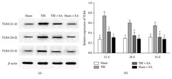
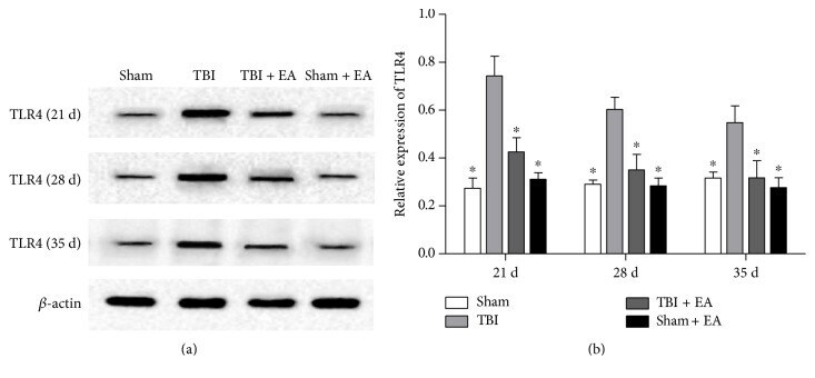
- Figure 2 EA treatment inhibited the level of toll-like receptor 4 (TLR4) in the hippocampus after TBI. (a) Representative Western blot (WB) bands of hippocampal TLR4 expression in the sham, TBI, TBI + EA, and sham + EA groups ( n = 9 in each group) at 21, 28, and 35 days posttrauma. (b) Quantitative analysis suggested that hippocampal TLR4 protein significantly increased in the TBI group compared with the sham group. And EA treatment caused an evident suppression of TBI-induced TLR4 upregulation in the TBI + EA group compared with the TBI and sham + EA groups. * P < 0.05 versus the TBI group.
- Submitted by
- Invitrogen Antibodies (provider)
- Main image
- Experimental details
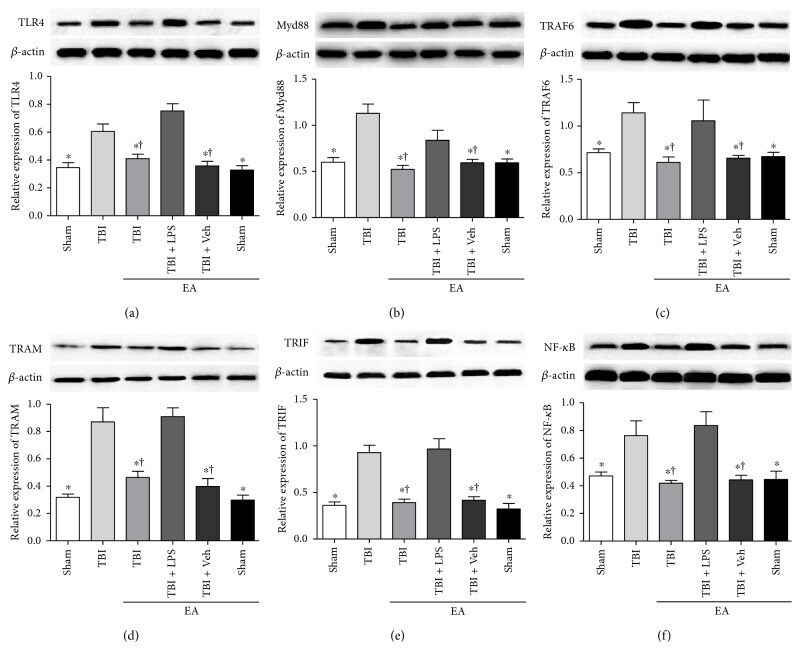
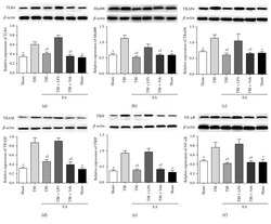
- Figure 5 EA treatment exerted an inhibitory effect on both of the two downstream pathways of TLR4 in the hippocampus posttrauma. Hippocampal TLR4, Myd88, TRAF6, TRAM, TRIF, and NF- kappa B expression of the sham, TBI, TBI + EA, TBI + EA + LPS, TBI + EA + Veh, and sham + EA groups ( n = 3 in each group) were detected by WB at 35 days after TBI. (a-f) The six kinds of proteins in TLR4/Myd88 kappa B and TLR4/TRIF/ kappa B signaling pathways were upregualted in the TBI group compared with the sham group and the sham + EA group. EA treatment resulted in a significant decrease in the TBI + EA group, but the effects were attenuated by LPS administration in the TBI + EA + LPS group. No difference of the proteins between the TBI + EA and TBI + EA + Veh groups was observed. * P < 0.05 versus the TBI group, + P < 0.05 versus the TBI + EA + LPS group.