 Explore
 Validate
 Learn
Learn Western blot
Antibody data
- Antibody Data
- Antigen structure
- References [0]
- Comments [0]
- Validations
- Western blot [2]
- Immunocytochemistry [1]
- Flow cytometry [2]
Submit
Validation data
Reference
Comment
Report error
- Product number
- 701363 - Provider product page

- Provider
- Invitrogen Antibodies
- Product name
- Phospho-S6 (Ser235, Ser236) Recombinant Rabbit Monoclonal Antibody (1H21L4)
- Antibody type
- Monoclonal
- Antigen
- Synthetic peptide
- Reactivity
- Human
- Host
- Rabbit
- Isotype
- IgG
- Antibody clone number
- 1H21L4
- Vial size
- 100 µg
- Concentration
- 0.5 mg/mL
- Storage
- Store at 4°C short term. For long term storage, store at -20°C, avoiding freeze/thaw cycles.
No comments: Submit comment
Supportive validation
- Submitted by
- Invitrogen Antibodies (provider)
- Main image
- Experimental details
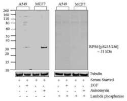
- Western blot analysis of RPS6 (pS235/236) was performed by loading 20 µg of Serum starved A549 (lane1), A459 treated for 10 minutes with 100 nM/mL of EGF (lane2), Serum starved MCF7 (Lane3) and MCF7 treated for 30 minutes with 25 µg/mL of Anisomycin cell lysates using Novex®NuPAGE®4-12 % Bis-Tris gel (Product # NP0321BOX), XCell SureLock Electrophoresis System (Product # EI0002), Novex® Sharp Pre-Stained Protein Standard (Product # LC5800), and iBlot® Dry Blotting System (Product # IB21001). Proteins were transferred to a nitrocellulose membrane and blocked with 5 % skim milk for 1 hour at room temperature. RPS6 (pS235/236) was detected at ~31 kDa using RPS6 (pS235/236) Recombinant Rabbit Monoclonal Antibody (Product # 701363) at 0.5-1 µg/mL in 2.5 % skim milk at 4°C overnight on a rocking platform. To confirm specificity, the corresponding blot on right was incubated with lambda phosphatase and its reactivity with antibody was tested. Goat anti-Rabbit IgG-HRP Secondary Antibody (Product # G-21234) at 1:5000 dilution was used and chemiluminescent detection was performed using Pierce™ ECL Western blotting Substrate (Product # 32106).

- Submitted by
- Invitrogen Antibodies (provider)
- Main image
- Experimental details
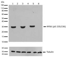
- Western blot analysis of Phospho-RPS6 pSer235/236 in whole cell extracts from HeLa treated with Calyculin A, HEK293 treated with Anisomycin, and serum-starved HeLa (20% FBS was added 4 hrs prior to lysing cells) (lanes 1, 3, 5 respectively) using a Phospho-RPS6 pSer235/236 recombinant rabbit monoclonal antibody (Product # 701363) at a dilution of 1 µg/mL. Tubulin was used as a loading control and detected with an anti-tubulin antibody. Samples were detected using chemiluminescence (ECL). Results show a band at ~31kDa.

Supportive validation
- Submitted by
- Invitrogen Antibodies (provider)
- Main image
- Experimental details
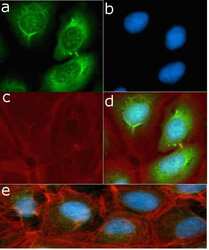
- Immunofluorescent analysis of Phospho-RPS6 pSer235/236 in U2OS cells using a Phospho-RPS6 pSer235/236 recombinant rabbit monoclonal antibody (Product # 701363) followed by detection using an Alexa Fluor 488-conjugated goat anti-rabbit secondary antibody (green) (Image A). Nuclei were stained using DAPI (Image B) and actin stained with Alexa Fluor 594 phalloidin (red) (image C). Image D is a composite image showing cytoplasmic localization and Image E is a composite image of cells showing inhibition of antibody binding after competition with the phosphorylated peptide.

Supportive validation
- Submitted by
- Invitrogen Antibodies (provider)
- Main image
- Experimental details
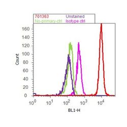
- Flow cytometry analysis of RPS6 [pS235/236] was done on HeLa cells. Cells were fixed with 70% ethanol for 10 minutes, permeabilized with 0.25% Tritonª X-100 for 20 minutes, and blocked with 5% BSA for 1 hour at room temperature. Cells were labeled with ABfinityª RPS6 [pS235/236] Recombinant Rabbit Monoclonal Antibody (701363, red histogram) or with rabbit isotype control (pink histogram) at 1-3 µg/million cells in 2.5% BSA. After incubation at room temperature for 2-3 hours, the cells were labeled with Alexa Fluor¨ 488 Goat Anti-Rabbit Secondary Antibody (A11008) at a dilution of 1:400 for 30 minutes at room temperature. The representative 10,000 cells were acquired and analyzed for each sample using an Attune¨ Acoustic Focusing Cytometer. The purple histogram represents unstained control cells and the green histogram represents no-primary-antibody control.

- Submitted by
- Invitrogen Antibodies (provider)
- Main image
- Experimental details
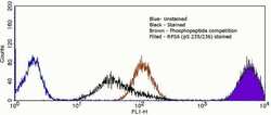
- Flow cytometry analysis of Phospho-RPS6 pSer235/236 in HeLa cells using a Phospho-RPS6 pSer235/236 recombinant rabbit monoclonal antibody (Product # 701363). Cells were fixed and permeabilized using FIX & PERM (Product # GAS-004) reagent, and detection was performed using an Alexa Fluor 488 goat anti-rabbit IgG (right peak) compared to an isotype control (middle peak, black) and a control without primary antibody (left peak, blue).
