 Explore
 Validate
 Learn
Learn Western blot
Antibody data
- Antibody Data
- Antigen structure
- References [0]
- Comments [0]
- Validations
- Western blot [3]
Submit
Validation data
Reference
Comment
Report error
- Product number
- 44-921G - Provider product page

- Provider
- Invitrogen Antibodies
- Product name
- Phospho-S6 (Ser236) Polyclonal Antibody
- Antibody type
- Polyclonal
- Antigen
- Synthetic peptide
- Reactivity
- Human
- Host
- Rabbit
- Isotype
- IgG
- Vial size
- 100 µL
- Storage
- -20°C
No comments: Submit comment
Supportive validation
- Submitted by
- Invitrogen Antibodies (provider)
- Main image
- Experimental details
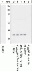
- Peptide Competition. Lysates prepared from HEK293 cells left untreated (1) or treated with EGF (2-6) were resolved by SDS-PAGE on a 14% polyacrylamide gel and transferred to PVDF. Membranes were blocked with a 5% BSA-TBST buffer for one hour at room temperature, and incubated with ribosomal protein S6 (pS236) antibody for two hours at room temperature in a 3% BSA-TBST buffer, following prior incubation with: no peptide (1, 2), the phosphopeptide corresponding to ribosomal protein S6 (pSpS244/247) (3), the non-phosphopeptide corresponding to the immunogen (4), a generic phosphoserine-containing peptide (5), or, the phosphopeptide immunogen (6). After washing, membranes were incubated with goat F (ab’)2 anti-rabbit IgG HRP conjugate (Product # ALI4404) and bands were detected using the Pierce SuperSignal™ method. The data show that only the peptide corresponding to ribosomal protein S6 (pS236) blocks the signal, verifying the specificity of the antibody.

- Submitted by
- Invitrogen Antibodies (provider)
- Main image
- Experimental details
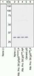
- Peptide Competition. Lysates prepared from HEK293 cells left untreated (1) or treated with EGF (2-6) were resolved by SDS-PAGE on a 14% polyacrylamide gel and transferred to PVDF. Membranes were blocked with a 5% BSA-TBST buffer for one hour at room temperature, and incubated with ribosomal protein S6 (pS236) antibody for two hours at room temperature in a 3% BSA-TBST buffer, following prior incubation with: no peptide (1, 2), the phosphopeptide corresponding to ribosomal protein S6 (pSpS244/247) (3), the non-phosphopeptide corresponding to the immunogen (4), a generic phosphoserine-containing peptide (5), or, the phosphopeptide immunogen (6). After washing, membranes were incubated with goat F (ab)2 anti-rabbit IgG HRP conjugate (Product # ALI4404) and bands were detected using the Pierce SuperSignal method. The data show that only the peptide corresponding to ribosomal protein S6 (pS236) blocks the signal, verifying the specificity of the antibody.

- Submitted by
- Invitrogen Antibodies (provider)
- Main image
- Experimental details
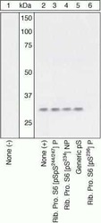
- Peptide Competition. Lysates prepared from HEK293 cells left untreated (1) or treated with EGF (2-6) were resolved by SDS-PAGE on a 14% polyacrylamide gel and transferred to PVDF. Membranes were blocked with a 5% BSA-TBST buffer for one hour at room temperature, and incubated with ribosomal protein S6 (pS236) antibody for two hours at room temperature in a 3% BSA-TBST buffer, following prior incubation with: no peptide (1, 2), the phosphopeptide corresponding to ribosomal protein S6 (pSpS244/247) (3), the non-phosphopeptide corresponding to the immunogen (4), a generic phosphoserine-containing peptide (5), or, the phosphopeptide immunogen (6). After washing, membranes were incubated with goat F (ab’)2 anti-rabbit IgG HRP conjugate (Product # ALI4404) and bands were detected using the Pierce SuperSignal™ method. The data show that only the peptide corresponding to ribosomal protein S6 (pS236) blocks the signal, verifying the specificity of the antibody.
