 Explore
 Validate
 Learn
Learn Western blot
Antibody data
- Antibody Data
- Antigen structure
- References [1]
- Comments [0]
- Validations
- Western blot [3]
- Flow cytometry [1]
Submit
Validation data
Reference
Comment
Report error
- Product number
- 44-922G - Provider product page

- Provider
- Invitrogen Antibodies
- Product name
- Phospho-S6 (Ser235, Ser236) Polyclonal Antibody
- Antibody type
- Polyclonal
- Antigen
- Synthetic peptide
- Reactivity
- Human
- Host
- Rabbit
- Isotype
- IgG
- Vial size
- 100 µL
- Storage
- -20°C
Submitted references Stimulation of autophagy by rapamycin protects neurons from remote degeneration after acute focal brain damage.
Viscomi MT, D'Amelio M, Cavallucci V, Latini L, Bisicchia E, Nazio F, Fanelli F, Maccarrone M, Moreno S, Cecconi F, Molinari M
Autophagy 2012 Feb 1;8(2):222-35
Autophagy 2012 Feb 1;8(2):222-35
No comments: Submit comment
Supportive validation
- Submitted by
- Invitrogen Antibodies (provider)
- Main image
- Experimental details
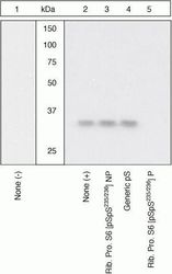
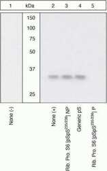
- Peptide Competition. Lysates prepared from HeLa cells left untreated (1) or treated with anisomycin (2-5). were resolved by SDS-PAGE on a 14% polyacrylamide gel and transferred to PVDF. Membranes were blocked with a 5% BSA-TBST buffer for one hour at room temperature, and incubated with ribosomal protein S6 (pSpS235/236) antibody for two hours at room temperature in a 3% BSA-TBST buffer, following prior incubation with: no peptide (1, 2), the non-phosphopeptide corresponding to the immunogen (3), a generic phosphoserine-containing peptide (4), or, the phosphopeptide immunogen (5). After washing, membranes were incubated with goat F (ab’)2 anti-rabbit IgG HRP conjugate (Product # ALI4404) and bands were detected using the Pierce SuperSignal™ method. The data show that only the peptide corresponding to ribosomal protein S6 (pSpS235/236) blocks the signal, verifying the specificity of the antibody.

- Submitted by
- Invitrogen Antibodies (provider)
- Main image
- Experimental details
- Peptide Competition. Lysates prepared from HeLa cells left untreated (1) or treated with anisomycin (2-5). were resolved by SDS-PAGE on a 14% polyacrylamide gel and transferred to PVDF. Membranes were blocked with a 5% BSA-TBST buffer for one hour at room temperature, and incubated with ribosomal protein S6 (pSpS235/236) antibody for two hours at room temperature in a 3% BSA-TBST buffer, following prior incubation with: no peptide (1, 2), the non-phosphopeptide corresponding to the immunogen (3), a generic phosphoserine-containing peptide (4), or, the phosphopeptide immunogen (5). After washing, membranes were incubated with goat F (ab’)2 anti-rabbit IgG HRP conjugate (Product # ALI4404) and bands were detected using the Pierce SuperSignal™ method. The data show that only the peptide corresponding to ribosomal protein S6 (pSpS235/236) blocks the signal, verifying the specificity of the antibody.

- Submitted by
- Invitrogen Antibodies (provider)
- Main image

- Experimental details
- Western blot analysis was performed on whole cell extracts (20 µg lysate) of HeLa (Lane 1), HeLa treated for 20 minutes with 200 nM of PMA (Lane 2), A549 (lane 3), Serum Starved A549 (lane 4) and A549 Serum Starved for overnight followed by Serum Released (lane 5). The blots were probed with Anti-Ribosomal Protein S6 (pS235)/(pS236) Rabbit Polyclonal Antibody (Product # 44-922G, 1:500 dilution) and detected by chemiluminescence using Goat anti-Rabbit IgG (H+L) Superclonal™ Secondary Antibody, HRP conjugate (Product # A27036, 0.4 µg/mL, 1:2500 dilution). A 31 kDa band corresponding to Ribosomal Protein S6 (Ser235/Ser236) were observed across PMA treated and Serum Starved followed by Serum Released cell lines tested. Known quantity of protein samples were electrophoresed using Novex® NuPAGE® 10 % Bis-Tris gel (Product # NP0302BOX), XCell SureLock™ Electrophoresis System (Product # EI0002) and Novex® Sharp Pre-Stained Protein Standard (Product # LC5800). Resolved proteins were then transferred onto a nitrocellulose membrane with iBlot® 2 Dry Blotting System (Product # IB21001). The membrane was probed with the relevant primary and secondary Antibody following blocking with 5 % skimmed milk. Chemiluminescent detection was performed using Pierce™ ECL Western Blotting Substrate (Product # 32106).

Supportive validation
- Submitted by
- Invitrogen Antibodies (provider)
- Main image

- Experimental details
- Flow cytometry analysis of Ribosomal Protein S6 [pS235/pS236] was done on HeLa cells. Cells were fixed with 70% ethanol for 10 minutes, permeabilized with 0.25% Triton™ X-100 for 20 minutes, and blocked with 5% BSA for 30 minutes at room temperature. Cells were labeled with Ribosomal Protein S6 [pS235/pS236] Rabbit Polyclonal Antibody (44922G, red histogram) or with rabbit isotype control (pink histogram) at 3-5 ug/million cells in 2.5% BSA. After incubation at room temperature for 2 hours, the cells were labeled with Alexa Fluor® 488 Goat Anti-Rabbit Secondary Antibody (A11008) at a dilution of 1:400 for 30 minutes at room temperature. The representative 10,000 cells were acquired and analyzed for each sample using an Attune® Acoustic Focusing Cytometer. The purple histogram represents unstained control cells and the green histogram represents no-primary-antibody control.
