 Explore
 Validate
 Learn
Learn Flow cytometry
Antibody data
- Antibody Data
- Antigen structure
- References [0]
- Comments [0]
- Validations
- Flow cytometry [1]
Submit
Validation data
Reference
Comment
Report error
- Product number
- 48-9007-41 - Provider product page

- Provider
- Invitrogen Antibodies
- Product name
- Anti-Phospho-S6 (Ser235, Ser236) Monoclonal Antibody (cupk43k), eFluor 450, eBioscience™
- Antibody type
- Monoclonal
- Antigen
- Other
- Description
- Description: This cupk43k monoclonal antibody recognizes human and mouse ribosomal protein S6 (also known as 40S ribosomal protein S6, phosphoprotein NP33, RPS6, RS6, S6) when phosphorylated on serine 235 (S235, human) and serine 236 (S236, mouse). Ribosomal protein S6 is a component of the 40S subunit of the ribosome and is phosphorylated at multiple sites following stimulation of cells by growth factors, tumor promoting agents, or mitogens. Phosphorylation of ribosomal protein S6 by p70S6K and PKDCD results in upregulation of the translation of RNA coding for proteins involved in cell cycle entry. Ribosomal protein S6 is dephosphorylated upon growth arrest. The specificity of the cupk43k monoclonal antibody was determined by western blotting. Applications Reported: This cupk43k antibody has been reported for use in intracellular staining followed by flow cytometric analysis. Applications Tested: This cupk43k antibody has been pre-titrated and tested by intracellular staining and flow cytometric analysis of stimulated normal human peripheral blood cells. This can be used at 5 µL (0.125 µg) per test. A test is defined as the amount (µg) of antibody that will stain a cell sample in a final volume of 100 µL. Cell number should be determined empirically but can range from 10^5 to 10^8 cells/test. Use of Protocol A: Two-step protocol: intracellular (cytoplasmic) proteins allows for the greatest flexibility for detection of surface and intracellular (cytoplasmic) proteins. Use of Protocol B: One-step protocol: intracellular (nuclear) proteins is recommended for staining of transcription factors in conjunction with surface and phosphorylated intracellular (cytoplasmic) proteins. Protocol C: Two-step protocol: Fixation/Methanol allows for the greatest discrimination of phospho-specific signaling between unstimulated and stimulated samples, but with limitations on the ability to stain specific surface proteins (refer to "Clone Performance Following Fixation/Permeabilization" located in the Best Protocols Section under the Resources tab online). All Protocols can be found in the "Staining Intracellular Antigens for Flow Cytometry Protocol" located in the Best Protocols Section under the Resources tab online. eFluor® 450 is an alternative to Pacific Blue®. eFluor® 450 emits at 445 nm and is excited with the Violet laser (405 nm). Please make sure that your instrument is capable of detecting this fluorochome. Excitation: 405 nm; Emission: 445 nm; Laser: Violet Laser. Filtration: 0.2 µm post-manufacturing filtered.
- Reactivity
- Human, Mouse
- Host
- Mouse
- Isotype
- IgG
- Antibody clone number
- cupk43k
- Vial size
- 25 Tests
- Concentration
- 5 µL/Test
- Storage
- 4° C, store in dark, DO NOT FREEZE!
No comments: Submit comment
Supportive validation
- Submitted by
- Invitrogen Antibodies (provider)
- Main image
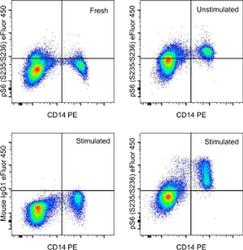
- Experimental details
- Intracellular staining of freshly-harvested (top left), unstimulated (top right), or 30-minute LPS-stimulated (bottom) normal human peripheral blood cells with Anti-Human CD14 PE (Product # 12-0149-42) and Mouse IgG1 K Isotype Control eFluor® 450 (Product # 48-4714-82) (bottom left) or Anti-Human phospho-S6 (S235/S236) APC (bottom right), using the Intracellular Fixation and Permeabilization Buffer Set (Product # 88-8824-00) and protocol. Cells in the lymphocyte/monocyte gate were used for analysis.
