Explore
 Validate
 Learn
Learn Western blot
 Immunocytochemistry
Antibody data
- Antibody Data
- Antigen structure
- References [2]
- Comments [0]
- Validations
- Immunocytochemistry [2]
- Immunohistochemistry [1]
- Flow cytometry [2]
- Other assay [1]
Submit
Validation data
Reference
Comment
Report error
- Product number
- MA5-15123 - Provider product page

- Provider
- Invitrogen Antibodies
- Product name
- S6 Monoclonal Antibody (C.896.4)
- Antibody type
- Monoclonal
- Antigen
- Recombinant full-length protein
- Description
- It is not recommended to aliquot this antibody.
- Reactivity
- Human, Mouse, Rat, Drosophila
- Host
- Mouse
- Isotype
- IgG
- Antibody clone number
- C.896.4
- Vial size
- 100 μL
- Concentration
- 5 μg/mL
- Storage
- -20°C
Submitted references Impaired dynamic interaction of axonal endoplasmic reticulum and ribosomes contributes to defective stimulus-response in spinal muscular atrophy.
Development of a Specific Monoclonal Antibody to Detect Male Cells Expressing the RPS4Y1 Protein.
Deng C, Reinhard S, Hennlein L, Eilts J, Sachs S, Doose S, Jablonka S, Sauer M, Moradi M, Sendtner M
Translational neurodegeneration 2022 Jun 2;11(1):31
Translational neurodegeneration 2022 Jun 2;11(1):31
Development of a Specific Monoclonal Antibody to Detect Male Cells Expressing the RPS4Y1 Protein.
Spena S, Cordiglieri C, Garagiola I, Peyvandi F
International journal of molecular sciences 2021 Feb 18;22(4)
International journal of molecular sciences 2021 Feb 18;22(4)
No comments: Submit comment
Supportive validation
- Submitted by
- Invitrogen Antibodies (provider)
- Main image

- Experimental details
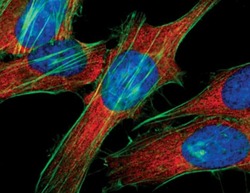
- Immunofluorescent analysis of S6 Ribosomal Protein in HeLa cells using a S6 Ribosomal Protein monoclonal antibody (Product # MA5-15123) (red). Actin filaments have been labeled with FITC-conjugated phalloidin. DNA is labeled using a fluorescent blue dye.

- Submitted by
- Invitrogen Antibodies (provider)
- Main image
- Experimental details
- Immunofluorescent analysis of S6 Ribosomal Protein in HeLa cells using a S6 Ribosomal Protein monoclonal antibody (Product # MA5-15123) (red). Actin filaments have been labeled with FITC-conjugated phalloidin. DNA is labeled using a fluorescent blue dye.

Supportive validation
- Submitted by
- Invitrogen Antibodies (provider)
- Main image
- Experimental details
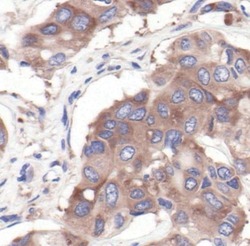
- Immunohistochemical analysis of S6 Ribosomal Protein in paraffin-embedded human lung carcinoma using a S6 Ribosomal Protein monoclonal antibody (Product # MA5-15123).

Supportive validation
- Submitted by
- Invitrogen Antibodies (provider)
- Main image

- Experimental details
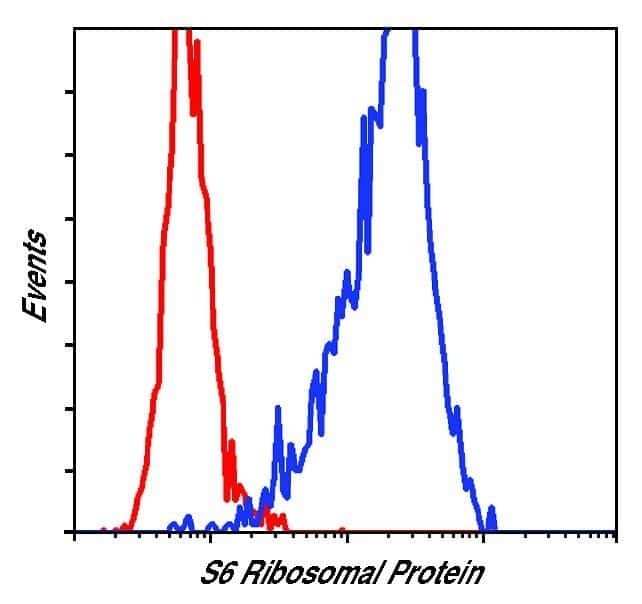
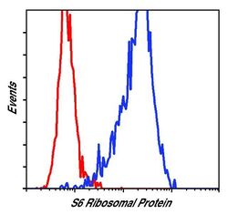
- Flow cytometric analysis of S6 Ribosomal Protein in NIH/3T3 cells using a S6 Ribosomal Protein monoclonal antibody (Product # MA5-15123) (blue) compared to a nonspecifc negative control antibody (red).
- Submitted by
- Invitrogen Antibodies (provider)
- Main image
- Experimental details
- Flow cytometric analysis of S6 Ribosomal Protein in NIH/3T3 cells using a S6 Ribosomal Protein monoclonal antibody (Product # MA5-15123) (blue) compared to a nonspecifc negative control antibody (red).

Supportive validation
- Submitted by
- Invitrogen Antibodies (provider)
- Main image
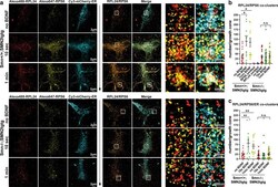
- Experimental details
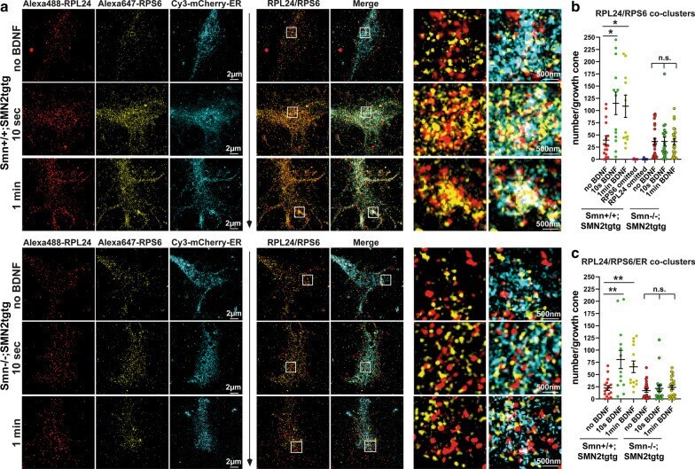
- Ribosome responsiveness to the BDNF/TrkB signaling as well as ribosome/ER tethering are impaired in Smn-deficient axon terminals. a BDNF-stimulated Smn + / + ;SMN2tgtg and Smn -/- ;SMN2tgtg motoneurons expressing mCherry-KDEL were stained against RPL24, RPS6 and mCherry-KDEL. Growth cones were imaged by SIM. White boxes indicate enlarged ROIs within growth cones. In control but not Smn -/- ;SMN2tgtg motoneurons, BDNF induces formation of RPL24/RPS6 co-clusters that colocalize with the axonal ER at 10 s and 1-min poststimulation. b Smn -/- ;SMN2tgtg motoneurons do not exhibit increased number of RPL24/RPS6 co-clusters upon stimulation (n.s., P = 0.9999; n = 21-23 cells), in contrast to Smn + / + ;SMN2tgtg (* P = 0.0147 for no BDNF vs. 10-s BDNF; * P = 0.0210 for no BDNF vs. 1-min BDNF; n = 13-16 cells from 3 independent experiments). In control experiments, either RPL24 or RPS6 antibodies were omitted to exclude the crosstalk between channels. Colocalization analysis detected almost no co-clusters in the growth cones of control neurons (see also Additional file 1 : Fig. S3). c In Smn + / + ;SMN2tgtg motoneurons, the number of RPL24/RPS6 co-clusters that colocalize with ER increases significantly within 10 s and 1-min of stimulation (** P = 0.0089 for no BDNF vs. 10 s BDNF; ** P = 0.0092 for no BDNF vs. 1-min BDNF; n = 13-16 cells from 3 independent experiments). In Smn -/- ;SMN2tgtg motoneurons the number of RPL24/RPS6 co-clusters that associate with ER does not increase upon