 Explore
 Validate
 Learn
Learn Western blot
Antibody data
- Antibody Data
- Antigen structure
- References [0]
- Comments [0]
- Validations
- Western blot [1]
- Immunocytochemistry [2]
- Immunohistochemistry [1]
Submit
Validation data
Reference
Comment
Report error
- Product number
- PA1-39503 - Provider product page

- Provider
- Invitrogen Antibodies
- Product name
- Phospho-S6 (Ser240, Ser244) Polyclonal Antibody
- Antibody type
- Polyclonal
- Antigen
- Synthetic peptide
- Description
- Heat-mediated antigen retrieval is recommended prior to staining, using a 10mM citrate buffer, pH 6.0, for 10 minutes followed by cooling at room temperature for 20 min. Following antigen retrieval, incubate samples with primary antibody for 30 min at room temperature. A suggested positive control is breast carcinoma. This antibody was originally validated as part of a Thermo Scientific Cellomics High Content Screening Kit. The antibody sold separately may have slightly different performance and may need to be further optimized for the best results.
- Reactivity
- Human
- Host
- Rabbit
- Isotype
- IgG
- Vial size
- 1 mL
- Storage
- Store at 4°C short term. For long term storage, store at -20°C, avoiding freeze/thaw cycles.
No comments: Submit comment
Supportive validation
- Submitted by
- Invitrogen Antibodies (provider)
- Main image

- Experimental details
- Western blot analysis was performed on whole cell extracts (30 µg lysate) of U-2 OS (Lane 1), U-2 OS treated with IGF (50 ng/mL for 6 hr) (Lane 2), U-2 OS treated with Rapamycin (50 nM for 4 hr) followed by treatment with IGF (50 ng/mL for 6hr) (Lane 3), A-431 (Lane 4), A431 treated with EGF (200 ng/mL for 10 min) and A-431 treated with Rapamycin (50 nM for 4 hr) followed by EGF treatment (200 ng/mL for 10 min). The blot was probed with Anti- Phospho-S6 (Ser240, Ser244) Monoclonal Antibody (Product # PA1-39503, 1:500 dilution) and detected by chemiluminescence using Goat anti-Rabbit IgG (H+L) Superclonal™ Secondary Antibody, HRP conjugate (Product # A27036, 0.25 µg/mL, 1:4000 dilution). A 32 kDa band corresponding to Phospho-S6 (Ser235, Ser236) was enhanced upon IGF and EGF treatment and was decreased upon rapamycin treatment on the cell lines tested.

Supportive validation
- Submitted by
- Invitrogen Antibodies (provider)
- Main image
- Experimental details
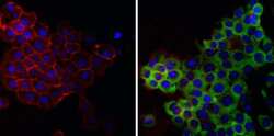
- Immunofluorescent analysis of Phospho-S6 pSer240/244 (green) in HeLa cells. Formalin fixed cells were permeabilized with 0.1% Triton X-100 in TBS for 10 minutes at room temperature and blocked with 1% Blocker BSA (Product # 37525) for 15 minutes at room temperature. Cells were probed without (left panel) or with (right panel) a Phospho-S6 pSer240/244 polyclonal antibody (Product # PA1-39503) at a dilution of 1:100 for at least 1 hour at room temperature, washed with PBS, and incubated with DyLight 488 goat anti-rabbit IgG secondary antibody (Product # 35552) at a dilution of 1:400 for 30 minutes at room temperature. F-Actin (red) was stained with DyLight 554 Phalloidin (Product # 21834) and nuclei (blue) were stained with Hoechst 33342 dye (Product # 62249). Images were taken on a Thermo Scientific ArrayScan or a ToxInsight Instrument at 20X magnification.

- Submitted by
- Invitrogen Antibodies (provider)
- Main image

- Experimental details
- Immunofluorescence analysis of Phospho-S6 (Ser240,Ser244) was performed using 70% confluent log phase U-2 OS cells treated with 50 ng/mL IGF-1 for 6hrs. The cells were fixed with 4% paraformaldehyde for 10 minutes, permeabilized with 0.1% Triton™ X-100 for 10 minutes, and blocked with 1% BSA for 1 hour at room temperature. The cells were labeled with Phospho-S6 (Ser240,Ser244) Polyclonal Antibody (Product # PA1-39503) at 1:100 dilution in 0.1% BSA, incubated overnight at 4 degree Celsius and then labeled with Goat anti-Rabbit IgG (H+L) Superclonal™ Secondary Antibody, Alexa Fluor® 488 conjugate (Product # A27034) at a dilution of 1:2000 for 45 minutes at room temperature (Panel a: green). Nuclei (Panel b: blue) were stained with SlowFade® Gold Antifade Mountant with DAPI (Product # S36938). F-actin (Panel c: red) was stained with Rhodamine Phalloidin (Product # R415, 1:300). Panel d represents the merged image showing cytoplasmic localization. Panel e represents cells treated with antagonist, Rapamycin (50 nM for 4hrs) followed by IGF (50 ng/mL for 6hrs), showing reduced expression of phospho-S6. Panel f shows the untreated cells showing basal expression of phospho S6. Panel g shows control cells with no primary antibody to assess background. The images were captured at 60X magnification.

Supportive validation
- Submitted by
- Invitrogen Antibodies (provider)
- Main image

- Experimental details
- Immunohistochemical analysis of Phospho-S6 Ribosomal pSer240/244 using anti-Phospho-S6 Ribosomal pSer240/244 Polyclonal Antibody (Product # PA5-32581) in Breast Carcinoma Cancer Tissue. The recommened dilution for this antibody in immunohistochemistry applications is 1:50.
