 Explore
 Validate
 Learn
Learn Western blot
 Immunocytochemistry
 Flow cytometry
Antibody data
- Antibody Data
- Antigen structure
- References [6]
- Comments [0]
- Validations
- Immunocytochemistry [1]
- Other assay [5]
Submit
Validation data
Reference
Comment
Report error
- Product number
- 44-1047G - Provider product page

- Provider
- Invitrogen Antibodies
- Product name
- Phospho-VEGF Receptor 2 (Tyr1054, Tyr1059) Polyclonal Antibody
- Antibody type
- Polyclonal
- Antigen
- Synthetic peptide
- Description
- On western blotting this antibody detects a protein with the apparent MW of 150 kDa. Positive controls used Porcine aortic endothelial (PAE) cells transfected with a chimeric receptor consisting of the extracellular domain of the CSF-1 receptor coupled to the transmembrane and cytoplasmic domains of the mouse VEGFR2, or 293T cells transfected with full length VEGFR2 and stimulated with 10 ng/mL VEGF for 5 minutes. VEGFR3 has not been tested, but is expected to react with this antibody.
- Reactivity
- Human, Mouse
- Host
- Rabbit
- Isotype
- IgG
- Vial size
- 100 µL
- Storage
- -20°C
Submitted references VLA-4 phosphorylation during tumor and immune cell migration relies on its coupling to VEGFR2 and CXCR4 by syndecan-1.
Epidermal-specific deletion of TC-PTP promotes UVB-induced epidermal cell survival through the regulation of Flk-1/JNK signaling.
Heparanase-induced shedding of syndecan-1/CD138 in myeloma and endothelial cells activates VEGFR2 and an invasive phenotype: prevention by novel synstatins.
Intramembrane binding of VE-cadherin to VEGFR2 and VEGFR3 assembles the endothelial mechanosensory complex.
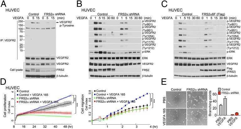
The docking protein FRS2α is a critical regulator of VEGF receptors signaling.
Investigation of the potential utility of a linomide analogue for treatment of choroidal neovascularization.
Jung O, Beauvais DM, Adams KM, Rapraeger AC
Journal of cell science 2019 Oct 28;132(20)
Journal of cell science 2019 Oct 28;132(20)
Epidermal-specific deletion of TC-PTP promotes UVB-induced epidermal cell survival through the regulation of Flk-1/JNK signaling.
Baek M, Kim M, Lim JS, Morales LD, Hernandez J, Mummidi S, Williams-Blangero S, Jang IS, Tsin AT, Kim DJ
Cell death & disease 2018 Jun 28;9(7):730
Cell death & disease 2018 Jun 28;9(7):730
Heparanase-induced shedding of syndecan-1/CD138 in myeloma and endothelial cells activates VEGFR2 and an invasive phenotype: prevention by novel synstatins.
Jung O, Trapp-Stamborski V, Purushothaman A, Jin H, Wang H, Sanderson RD, Rapraeger AC
Oncogenesis 2016 Feb 29;5(2):e202
Oncogenesis 2016 Feb 29;5(2):e202
Intramembrane binding of VE-cadherin to VEGFR2 and VEGFR3 assembles the endothelial mechanosensory complex.
Coon BG, Baeyens N, Han J, Budatha M, Ross TD, Fang JS, Yun S, Thomas JL, Schwartz MA
The Journal of cell biology 2015 Mar 30;208(7):975-86
The Journal of cell biology 2015 Mar 30;208(7):975-86
The docking protein FRS2α is a critical regulator of VEGF receptors signaling.
Chen PY, Qin L, Zhuang ZW, Tellides G, Lax I, Schlessinger J, Simons M
Proceedings of the National Academy of Sciences of the United States of America 2014 Apr 15;111(15):5514-9
Proceedings of the National Academy of Sciences of the United States of America 2014 Apr 15;111(15):5514-9
Investigation of the potential utility of a linomide analogue for treatment of choroidal neovascularization.
Abdel-Rahman MH, Yang Y, Salem MM, Meadows S, Massengill JB, Li PK, Davidorf FH
Experimental eye research 2010 Dec;91(6):837-43
Experimental eye research 2010 Dec;91(6):837-43
No comments: Submit comment
Supportive validation
- Submitted by
- Invitrogen Antibodies (provider)
- Main image
- Experimental details
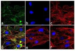
- Immunofluorescence analysis of Phospho-VEGF Receptor 2 pTyr1054/pTyr1059 was done on 90% confluent log phase HUVEC cell treated with 100 ng of VEGF for 30 minutes. The cells were fixed with 4% paraformaldehyde for 10 minutes, permeabilized with 0.1% Triton™ X-100 for 10 minutes, and blocked with 1% BSA for 1 hour at room temperature. The cells were labeled with Phospho-VEGF Receptor 2 pTyr1054/pTyr1059 Rabbit Polyclonal Antibody (Product # 44-1047G) at 1:250 dilution in 0.1% BSA and incubated for 3 hours at room temperature and then labeled with Goat anti-Rabbit IgG (H+L) Superclonal™ Secondary Antibody, Alexa Fluor® 488 conjugate (Product # A27034) at a dilution of 1:2000 for 45 minutes at room temperature (Panel a: green). Nuclei (Panel b: blue) were stained with SlowFade® Gold Antifade Mountant with DAPI (Product # S36938). F-actin (Panel c: red) was stained with Rhodamine Phalloidin (Product # R415, 1:300). Panel d is a merged image showing punctuated membranous localization. Panel e is untreated cell with no signal. Panel f is a no primary antibody control. The images were captured at 60X magnification.
Supportive validation
- Submitted by
- Invitrogen Antibodies (provider)
- Main image
- Experimental details
- NULL
- Submitted by
- Invitrogen Antibodies (provider)
- Main image
- Experimental details
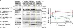
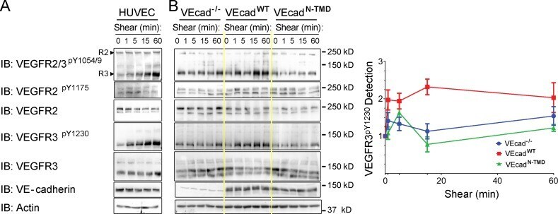
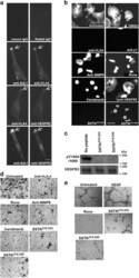
- Figure 4. VEcad TMD in shear-mediated VEGFR2 and VEGFR3 activation. (A) VEGFR activation. HUVECs exposed to 12 dynes/cm 2 laminar shear for the indicated times were immunoblotted with the indicated antibodies. The upper band on the anti-VEGFR2/3 pY1054/9 blot is active VEGFR2; the lower band is active VEGFR3 (arrowheads). IB, immunoblotting. (B) Dependence on TMD. VEcad -/- , VEcad WT , and VEcad DeltaTMD cells were subjected to shear stress, then lysed and immunoblotted as in A. The anti-VEGFR pY1230 blots were quantified by densitometry of the ~120-kD band with actin serving as a loading control. Values are means +- SEM (error bars), n >= 3. Yellow lines mark boundaries between cell types.
- Submitted by
- Invitrogen Antibodies (provider)
- Main image
- Experimental details
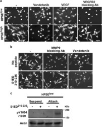
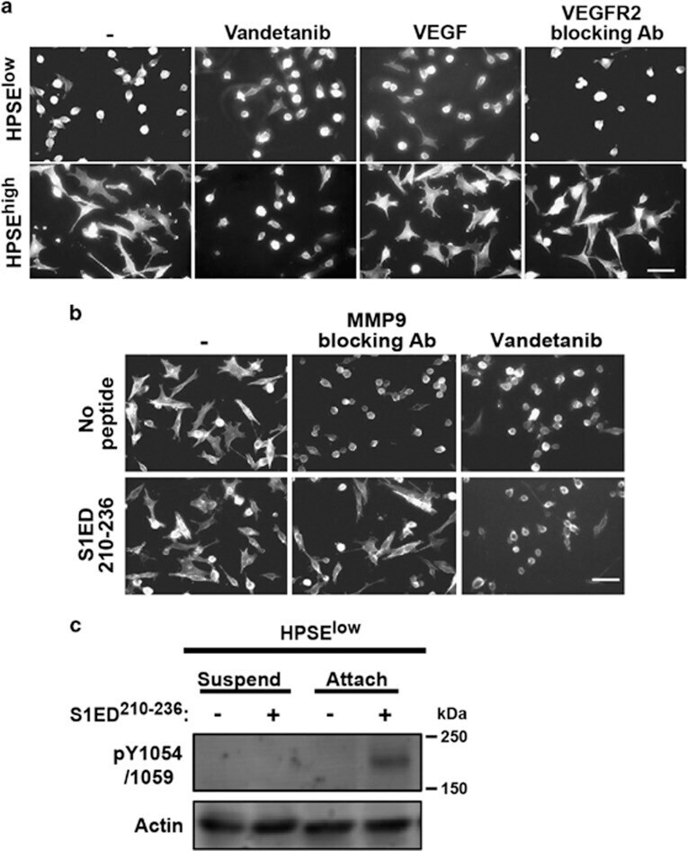
- Figure 4 HPSE causes activation of VEGFR2 when VLA-4 engages ligand. ( a ) HPSE low and HPSE high cells treated with or without 1 mu M Vandetanib, 20 ng/ml VEGF or 20 mug/ml VEGFR2 blocking antibody are plated on VCAM-1 to observe cell spreading (bar=50 mum). ( b ) HPSE high cells plated on FN were treated with MMP-9 blocking antibody or 1 mu M Vandetanib in the absence or presence of 0.3 mu M S1ED 210-236 (bar=50 mum). ( c ) HPSE low cells were kept in suspension or were seeded on FN in the absence or presence of 0.3 mu M S1ED 210-236 for 2.5 h. Cell lysates were immunoblotted with antibodies against pY1054/1059 in VEGFR2 as a marker of VEGFR2 activation. Actin is shown as a loading control.
- Submitted by
- Invitrogen Antibodies (provider)
- Main image
- Experimental details
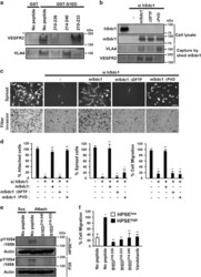
- Figure 6 SSTN peptides specific for VEGFR2 or VLA-4 compete with shed Sdc1 and inhibit the HPSE-induced invasive phenotype. ( a ) HPSE low cell lysates were incubated overnight with glutathione beads coated with GST or GST-S1ED in the absence or presence of 30 mu M S1ED 210-236 , S1ED 214-240 or S1ED 210-233 . Capture of VLA-4 or VEGFR2 was detected by immunoblotting. ( b - d ) HPSE high cells were untreated, transfected with hSdc1 small interfering RNA (siRNA) alone or transfected with with hSdc1 siRNA together with cDNA for mSdc1, mSdc1 DeltaDFTF or mSdc1 DeltaPVD for 72 h, before performing immunoprecipitation, cell spreading or migration assays. ( b ) Top: blots were probed for hSdc1, mSdc1 or mSdc1 mutants following immunoprecipitation from whole-cell lysates. Bottom: Sdc1 captured from the conditioned medium of each cell treatment by mouse-Sdc1-specific mAb281-2-coated beads was incubated with CAG cell lysates, and probed on blots for co-precipitation of VLA-4 or VEGFR2. ( c ) Top: pictures of cell attachment and spreading following plating on FN for 2.5 h (bar=50 mum). Bottom: pictures after 12-h migration through FN-coated filters (bar=100 mum). ( d ) Quantification of cell attachment or spreading on FN and cell migration toward FN. * P

- Submitted by
- Invitrogen Antibodies (provider)
- Main image
- Experimental details
- Figure 8 The Sdc1-coupled VEGFR2-VLA-4 complex induces angiogenesis in endothelial cells. ( a ) HMEC-1 cells were plated on the IIICS FN fragment for 5 h and double stained for VLA-4 and Sdc1, VEGFR2 and Sdc1, or VLA-4 and VEGFR2 (bar=10 mum). ( b ) HMEC-1 cells plated on IIICS were treated with DMSO vehicle, VLA-4 blocking antibody (P1H4), 10 mug/ml beta1-integrin blocking antibody mAb13, MMP-9 blocking antibody, Roneparstat (Rone) (125 mug/ml), Vandetanib, VEGFR2 blocking antibody, 30 mu M SSTN 210-233 or SSTN 214-240 (bar=50 mum). ( c ) HMEC-1 cells were plated on IIICS for 2 h in the presence of 10 mu M of SSTN 210-233 , or SSTN 214-240 . Lysates were probed on immunoblots with antibodies against p1054/1059 of VEGFR2 and total VEGFR2. Note that other intervening lanes in the blot were removed. ( d ) Sixteen-hour transfilter migration assays toward GST-IIICS were performed for HMEC-1 cells in the presence of VLA-4 blocking antibody, Roneparstat (Rone), MMP-9 blocking antibody, Vandetanib, or 30 mu M SSTN 210-233 or SSTN 214-240 (bar=100 mum). ( e ) HMEC-1 cells (2.5 x 10 4 cells per well) were seeded onto matrigel containing GST-IIICS and cultured in media containing 20 ng/ml VEGF in the absence or presence of Roneparstat (Rone), or 30 mu M of SSTN 210-233 , or SSTN 214-240 for 24 h. Images of random fields were taken at 100x to observe capillary network formation (bar=250 mum).
