Explore
 Validate
 Learn
Learn Western blot
 Immunocytochemistry
Antibody data
- Antibody Data
- Antigen structure
- References [2]
- Comments [0]
- Validations
- Immunocytochemistry [1]
Submit
Validation data
Reference
Comment
Report error
- Product number
- HPA019592 - Provider product page

- Provider
- Atlas Antibodies
- Proper citation
- Atlas Antibodies Cat#HPA019592, RRID:AB_10601499
- Product name
- Anti-S100A13
- Antibody type
- Polyclonal
- Description
- Polyclonal Antibody against Human S100A13, Gene description: S100 calcium binding protein A13, Validated applications: ICC, IHC, WB, Uniprot ID: Q99584, Storage: Store at +4°C for short term storage. Long time storage is recommended at -20°C.
- Reactivity
- Human, Rat
- Host
- Rabbit
- Conjugate
- Unconjugated
- Isotype
- IgG
- Vial size
- 100 µl
- Concentration
- 0.5 mg/ml
- Storage
- Store at +4°C for short term storage. Long time storage is recommended at -20°C.
- Handling
- The antibody solution should be gently mixed before use.
Submitted references Differential Protein Expression Profiles of Cyst Fluid from Papillary Thyroid Carcinoma and Benign Thyroid Lesions
Proteomics analysis of melanoma metastases: association between S100A13 expression and chemotherapy resistance
Dumont J, Dinets A, Pernemalm M, Kjellin H, Sviatoha V, Sofiadis A, Juhlin C, Zedenius J, Larsson C, Lehtiö J, Höög A
PLOS ONE 2015;10(5):e0126472
PLOS ONE 2015;10(5):e0126472
Proteomics analysis of melanoma metastases: association between S100A13 expression and chemotherapy resistance
Azimi A, Pernemalm M, Frostvik Stolt M, Hansson J, Lehtiö J, Egyházi Brage S, Hertzman Johansson C
British Journal of Cancer 2014;110(10):2489-2495
British Journal of Cancer 2014;110(10):2489-2495
No comments: Submit comment
Supportive validation
- Submitted by
- Atlas Antibodies (provider)
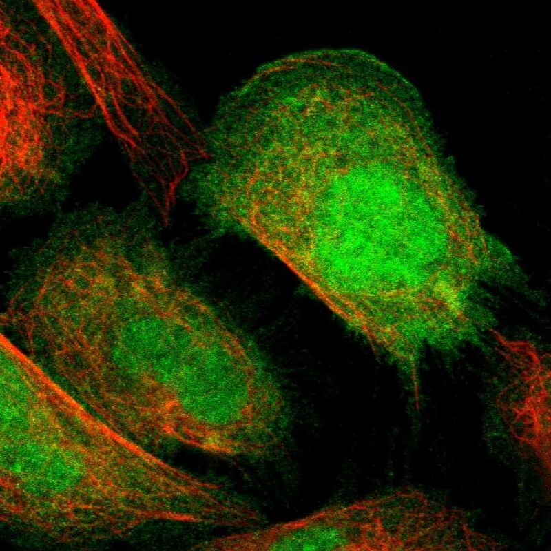
- Main image
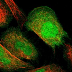
- Experimental details
- Immunofluorescent staining of human cell line U-2 OS shows localization to nucleus, plasma membrane & cytosol.
- Sample type
- Human