Explore
 Validate
 Learn
Learn Western blot
 Immunohistochemistry
Antibody data
- Antibody Data
- Antigen structure
- References [0]
- Comments [0]
- Validations
- Immunohistochemistry [2]
- Flow cytometry [1]
- Other assay [2]
Submit
Validation data
Reference
Comment
Report error
- Product number
- STJ72494 - Provider product page

- Provider
- St John's Laboratory
- Product name
- Anti-CRTC2 antibody (C-Term) (STJ72494)
- Antibody type
- Polyclonal
- Description
- Goat polyclonal antibody anti-CRTC2 (C-Term) is suitable for use in ELISA, Western Blot and Immunohistochemistry research applications.
- Reactivity
- Human, Mouse, Rat, Bovine, Canine, Porcine
- Host
- Goat
- Conjugate
- Unconjugated
- Antigen sequence
DPAVEDSFRSDRLQ- Epitope
- NA
- Isotype
- IgG
- Antibody clone number
- NA
- Vial size
- NA
- Concentration
- NA
- Storage
- Store at-20 on receipt and minimise freeze-thaw cycles.
- Handling
- NA
No comments: Submit comment
Supportive validation
Supportive validation
- Submitted by
- St John's Laboratory (provider)
- Main image
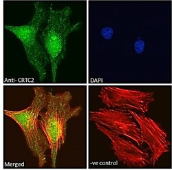
- Experimental details
- STJ72494 Immunofluorescence analysis of paraformaldehyde fixed HeLa cells, permeabilized with 0. 15% Triton. Primary incubation 1hr (10ug/ml) followed by Alexa Fluor 488 secondary antibody (2ug/ml) , showing strong nuclear staining. Actin filaments were stained with phalloidin (red) and the nuclear stain is DAPI (blue). Negative control: Unimmunized goat IgG (10ug/ml) followed by Alexa Fluor 488 secondary antibody (2ug/ml).
- Sample type
- NA
- Validation comment
- NA
- Primary Ab dilution
- NA
- Other comments
- NA
- Secondary Ab
- NA
- Secondary Ab dilution
- NA
- Protocol
- NA
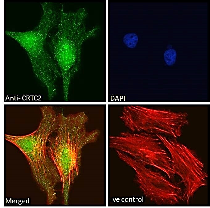
Supportive validation
- Submitted by
- St John's Laboratory (provider)
- Main image

- Experimental details
- STJ72494 Immunofluorescence analysis of paraformaldehyde fixed U2OS cells, permeabilized with 0. 15% Triton. Primary incubation 1hr (10ug/ml) followed by Alexa Fluor 488 secondary antibody (2ug/ml) , showing strong nuclear staining. Actin filaments were stained with phalloidin (red) and the nuclear stain is DAPI (blue). Negative control: Unimmunized goat IgG (10ug/ml) followed by Alexa Fluor 488 secondary antibody (2ug/ml).
- Sample type
- NA
- Validation comment
- NA
- Primary Ab dilution
- NA
- Other comments
- NA
- Secondary Ab
- NA
- Secondary Ab dilution
- NA
- Protocol
- NA
Supportive validation
- Submitted by
- St John's Laboratory (provider)
- Main image
- Experimental details
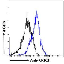
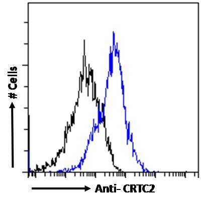
- STJ72494 Flow cytometric analysis of paraformaldehyde fixed HeLa cells (blue line) , permeabilized with 0. 5% Triton. Primary incubation 1hr (10ug/ml) followed by Alexa Fluor 488 secondary antibody (1ug/ml). IgG control: Unimmunized goat IgG (black line) followed by Alexa Fluor 488 secondary antibody.
- Sample type
- NA
- Validation comment
- NA
- Primary Ab dilution
- NA
- Other comments
- NA
- Secondary Ab
- NA
- Secondary Ab dilution
- NA
- Protocol
- NA
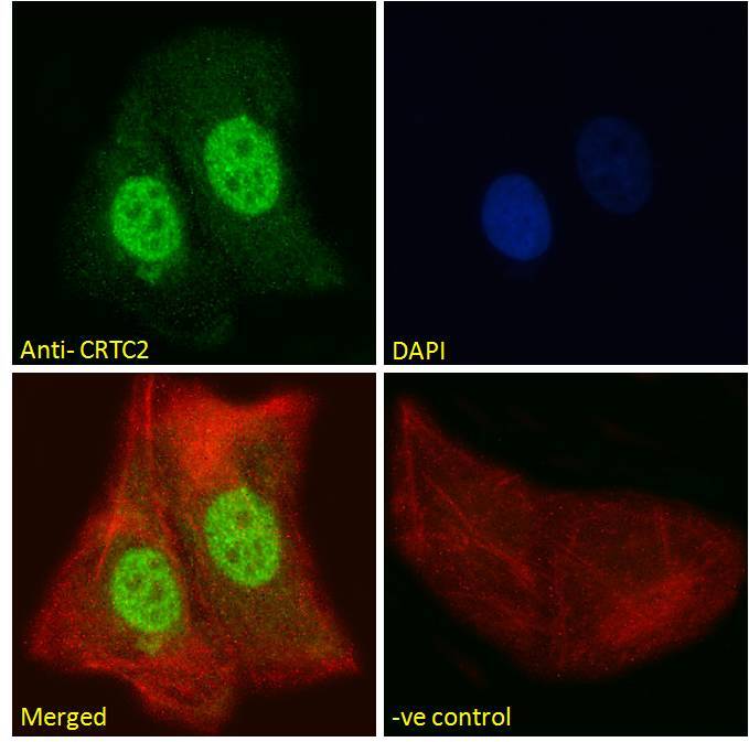
Supportive validation
Supportive validation
- Submitted by
- St John's Laboratory (provider)
- Main image

- Experimental details
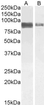
- STJ72494 (1µg/ml) staining of Jurkat (A) and (2ug/ml) A431 (B) cell lysate (35µg protein in RIPA buffer). Detected by chemiluminescence.
- Sample type
- NA
- Validation comment
- NA
- Primary Ab dilution
- NA
- Other comments
- NA
- Secondary Ab
- NA
- Secondary Ab dilution
- NA
- Protocol
- NA
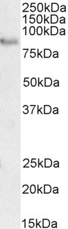
Supportive validation
- Submitted by
- St John's Laboratory (provider)
- Main image

- Experimental details
- STJ72494 (2µg/ml) staining of Rat Brain lysate (35µg protein in RIPA buffer). Detected by chemiluminescence.
- Sample type
- NA
- Validation comment
- NA
- Primary Ab dilution
- NA
- Other comments
- NA
- Secondary Ab
- NA
- Secondary Ab dilution
- NA
- Protocol
- NA