Explore
 Validate
 Learn
Learn51-3500
antibody from Invitrogen Antibodies
Targeting: NFKB1
KBF1, NF-kappaB, NF-kB1, NFkappaB, NFKB-p50, p105, p50
 Western blot
 Immunocytochemistry
Antibody data
- Antibody Data
- Antigen structure
- References [9]
- Comments [0]
- Validations
- Immunocytochemistry [2]
- Immunohistochemistry [3]
- Flow cytometry [1]
- Other assay [7]
Submit
Validation data
Reference
Comment
Report error
- Product number
- 51-3500 - Provider product page

- Provider
- Invitrogen Antibodies
- Product name
- NFkB p50 Polyclonal Antibody
- Antibody type
- Polyclonal
- Antigen
- Recombinant full-length protein
- Reactivity
- Human, Mouse
- Host
- Rabbit
- Isotype
- IgG
- Vial size
- 100 μg
- Concentration
- 0.25 mg/mL
- Storage
- -20°C
Submitted references Silencing of lncRNA XIST impairs angiogenesis and exacerbates cerebral vascular injury after ischemic stroke.
KLF4 alleviates cerebral vascular injury by ameliorating vascular endothelial inflammation and regulating tight junction protein expression following ischemic stroke.
Establishment of three novel cell lines derived from African American patients with colorectal carcinoma: A unique tool for assessing racial health disparity.
Photobiomodulation with Pulsed and Continuous Wave Near-Infrared Laser (810 nm, Al-Ga-As) Augments Dermal Wound Healing in Immunosuppressed Rats.
Superpulsed (Ga-As, 904 nm) low-level laser therapy (LLLT) attenuates inflammatory response and enhances healing of burn wounds.
Inhibition of rotavirus ECwt infection in ICR suckling mice by N-acetylcysteine, peroxisome proliferator-activated receptor gamma agonists and cyclooxygenase-2 inhibitors.
Human gut flora-fermented nondigestible fraction from cooked bean ( Phaseolus vulgaris L.) modifies protein expression associated with apoptosis, cell cycle arrest, and proliferation in human adenocarcinoma colon cancer cells.
Measles virus-induced modulation of host-cell gene expression.
Mediation of proliferating cell nuclear antigen (PCNA)-dependent DNA replication through a conserved p21(Cip1)-like PCNA-binding motif present in the third subunit of human DNA polymerase delta.
Wang C, Dong J, Sun J, Huang S, Wu F, Zhang X, Pang D, Fu Y, Li L
Molecular therapy. Nucleic acids 2021 Dec 3;26:148-160
Molecular therapy. Nucleic acids 2021 Dec 3;26:148-160
KLF4 alleviates cerebral vascular injury by ameliorating vascular endothelial inflammation and regulating tight junction protein expression following ischemic stroke.
Zhang X, Wang L, Han Z, Dong J, Pang D, Fu Y, Li L
Journal of neuroinflammation 2020 Apr 7;17(1):107
Journal of neuroinflammation 2020 Apr 7;17(1):107
Establishment of three novel cell lines derived from African American patients with colorectal carcinoma: A unique tool for assessing racial health disparity.
Paredes J, Ji P, Lacomb JF, Shroyer KR, Martello LA, Williams JL
International journal of oncology 2018 Oct;53(4):1516-1528
International journal of oncology 2018 Oct;53(4):1516-1528
Photobiomodulation with Pulsed and Continuous Wave Near-Infrared Laser (810 nm, Al-Ga-As) Augments Dermal Wound Healing in Immunosuppressed Rats.
Keshri GK, Gupta A, Yadav A, Sharma SK, Singh SB
PloS one 2016;11(11):e0166705
PloS one 2016;11(11):e0166705
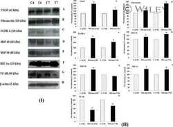
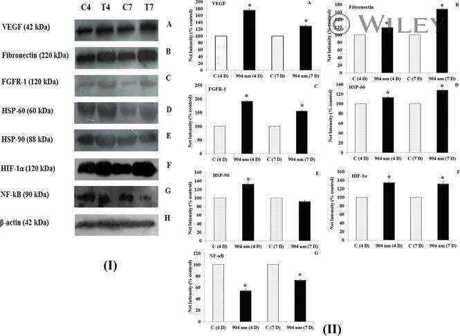
Superpulsed (Ga-As, 904 nm) low-level laser therapy (LLLT) attenuates inflammatory response and enhances healing of burn wounds.
Gupta A, Keshri GK, Yadav A, Gola S, Chauhan S, Salhan AK, Bala Singh S
Journal of biophotonics 2015 Jun;8(6):489-501
Journal of biophotonics 2015 Jun;8(6):489-501
Inhibition of rotavirus ECwt infection in ICR suckling mice by N-acetylcysteine, peroxisome proliferator-activated receptor gamma agonists and cyclooxygenase-2 inhibitors.
Guerrero CA, Paula Pardo VR, Rafael Guerrero OA
Memorias do Instituto Oswaldo Cruz 2013 Sep;108(6):741-54
Memorias do Instituto Oswaldo Cruz 2013 Sep;108(6):741-54
Human gut flora-fermented nondigestible fraction from cooked bean ( Phaseolus vulgaris L.) modifies protein expression associated with apoptosis, cell cycle arrest, and proliferation in human adenocarcinoma colon cancer cells.
Campos-Vega R, García-Gasca T, Guevara-Gonzalez R, Ramos-Gomez M, Oomah BD, Loarca-Piña G
Journal of agricultural and food chemistry 2012 Dec 26;60(51):12443-50
Journal of agricultural and food chemistry 2012 Dec 26;60(51):12443-50
Measles virus-induced modulation of host-cell gene expression.
Bolt G, Berg K, Blixenkrone-Møller M
The Journal of general virology 2002 May;83(Pt 5):1157-1165
The Journal of general virology 2002 May;83(Pt 5):1157-1165
Mediation of proliferating cell nuclear antigen (PCNA)-dependent DNA replication through a conserved p21(Cip1)-like PCNA-binding motif present in the third subunit of human DNA polymerase delta.
Ducoux M, Urbach S, Baldacci G, Hübscher U, Koundrioukoff S, Christensen J, Hughes P
The Journal of biological chemistry 2001 Dec 28;276(52):49258-66
The Journal of biological chemistry 2001 Dec 28;276(52):49258-66
No comments: Submit comment
Supportive validation
- Submitted by
- Invitrogen Antibodies (provider)
- Main image

- Experimental details
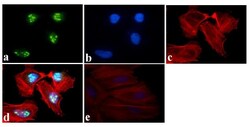
- Immunofluorescent analysis of NFkB p105/p50 Antibody was done on 70% confluent log phase HeLa cells. The cells were fixed with 4% paraformaldehyde for 15 minutes, permeabilized with 0.25% Triton™ X-100 for 10 minutes, and blocked with 5% BSA for 1 hour at room temperature. The cells were labeled with NFkB p105/p50 Antibody (Product # 38-5600) at 1µg/mL in 1% BSA and incubated for 3 hours at room temperature and then labeled with Alexa Fluor 488 Goat Anti-Rabbit IgG Secondary Antibody (Product # A-11008) at a dilution of 1:400 for 45 minutes at room temperature (Panel a: green). Nuclei (Panel b: blue) were stained with SlowFade® Gold Antifade Mountant with DAPI (Product # S36938). F-actin (Panel c: red) was stained with Alexa Fluor 594 Phalloidin (Product # A12381). Panel d is a merged image showing Nuclear localization. Panel e is a no primary antibody control. The images were captured at 40X magnification.

- Submitted by
- Invitrogen Antibodies (provider)
- Main image
- Experimental details
- Immunofluorescent analysis of NFkB p105/p50 Antibody was done on 70% confluent log phase HeLa cells. The cells were fixed with 4% paraformaldehyde for 15 minutes, permeabilized with 0.25% Triton™ X-100 for 10 minutes, and blocked with 5% BSA for 1 hour at room temperature. The cells were labeled with NFkB p105/p50 Antibody (Product # 38-5600) at 1µg/mL in 1% BSA and incubated for 3 hours at room temperature and then labeled with Alexa Fluor 488 Goat Anti-Rabbit IgG Secondary Antibody (Product # A-11008) at a dilution of 1:400 for 45 minutes at room temperature (Panel a: green). Nuclei (Panel b: blue) were stained with SlowFade® Gold Antifade Mountant with DAPI (Product # S36938). F-actin (Panel c: red) was stained with Alexa Fluor 594 Phalloidin (Product # A12381). Panel d is a merged image showing Nuclear localization. Panel e is a no primary antibody control. The images were captured at 40X magnification.

Supportive validation
- Submitted by
- Invitrogen Antibodies (provider)
- Main image
- Experimental details
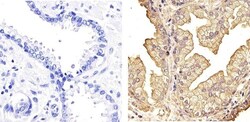
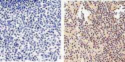
- Immunohistochemistry analysis of NF-kappa-B-p105/p50 showing staining in the nucleus and cytoplasm of paraffin-embedded human prostate tissue (right) compared to a negative control without primary antibody (left). To expose target proteins, antigen retrieval was performed using 10mM sodium citrate (pH 6.0), microwaved for 8-15 min. Following antigen retrieval, tissues were blocked in 3% H2O2-methanol for 15 min at room temperature, washed with ddH2O and PBS, and then probed with a NF-kappa-B-p105/p50 polyclonal antibody (Product # 51-3500) diluted in 3% BSA-PBS at a dilution of 1:100 overnight at 4°C in a humidified chamber. Tissues were washed extensively in PBST and detection was performed using an HRP-conjugated secondary antibody followed by colorimetric detection using a DAB kit. Tissues were counterstained with hematoxylin and dehydrated with ethanol and xylene to prep for mounting.
- Submitted by
- Invitrogen Antibodies (provider)
- Main image
- Experimental details
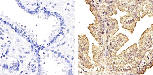
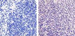
- Immunohistochemistry analysis of NF-kappa-B-p105/p50 showing staining in the nucleus and cytoplasm of paraffin-embedded human spleen tissue (right) compared to a negative control without primary antibody (left). To expose target proteins, antigen retrieval was performed using 10mM sodium citrate (pH 6.0), microwaved for 8-15 min. Following antigen retrieval, tissues were blocked in 3% H2O2-methanol for 15 min at room temperature, washed with ddH2O and PBS, and then probed with a NF-kappa-B-p105/p50 polyclonal antibody (Product # 51-3500) diluted in 3% BSA-PBS at a dilution of 1:100 overnight at 4°C in a humidified chamber. Tissues were washed extensively in PBST and detection was performed using an HRP-conjugated secondary antibody followed by colorimetric detection using a DAB kit. Tissues were counterstained with hematoxylin and dehydrated with ethanol and xylene to prep for mounting.

- Submitted by
- Invitrogen Antibodies (provider)
- Main image
- Experimental details
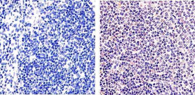
- Immunohistochemistry analysis of NF-kappa-B-p105/p50 showing staining in the nucleus and cytoplasm of paraffin-embedded mouse spleen tissue (right) compared to a negative control without primary antibody (left). To expose target proteins, antigen retrieval was performed using 10mM sodium citrate (pH 6.0), microwaved for 8-15 min. Following antigen retrieval, tissues were blocked in 3% H2O2-methanol for 15 min at room temperature, washed with ddH2O and PBS, and then probed with a NF-kappa-B-p105/p50 polyclonal antibody (Product # 51-3500) diluted in 3% BSA-PBS at a dilution of 1:20 overnight at 4°C in a humidified chamber. Tissues were washed extensively in PBST and detection was performed using an HRP-conjugated secondary antibody followed by colorimetric detection using a DAB kit. Tissues were counterstained with hematoxylin and dehydrated with ethanol and xylene to prep for mounting.
Supportive validation
- Submitted by
- Invitrogen Antibodies (provider)
- Main image
- Experimental details
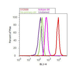
- Flow cytometry analysis of NFkB was done on HeLa cells. Cells were fixed with 70% ethanol for 10 minutes, permeabilized with 0.25% Triton™ X-100 for 20 minutes, and blocked with 5% BSA for 30 minutes at room temperature. Cells were labeled with NFkB Rabbit Polyclonal Antibody (513500, red histogram) or with rabbit isotype control (pink histogram) at 3-5 ug/million cells in 2.5% BSA. After incubation at room temperature for 2 hours, the cells were labeled with Alexa Fluor® 488 Goat Anti-Rabbit Secondary Antibody (A11008) at a dilution of 1:400 for 30 minutes at room temperature. The representative 10,000 cells were acquired and analyzed for each sample using an Attune® Acoustic Focusing Cytometer. The purple histogram represents unstained control cells and the green histogram represents no-primary-antibody control.

Supportive validation
- Submitted by
- Invitrogen Antibodies (provider)
- Main image
- Experimental details
- NULL
- Submitted by
- Invitrogen Antibodies (provider)
- Main image

- Experimental details
- Fig. 5: increased expression of cyclooxygenase (COX)-2, ERp57, Hsc70, nuclearfactor kappa B (NF-IoB), Hsp70, protein disulphide isomerase (PDI) andperoxisome proliferator-activated receptor gamma (PPARI3) in villuscells infected by ECwt. Mice were infected with ECwt and at thre dayspost-inoculation (d.p.i.) their small intestinal villi were isolated.Villi were analysed by immunocytochemistry (A) or flow cytometry (B) (n= 2 mice for each analysis). A: the immunocytochemistry analysis ofvillus cross-sections was performed by using rabbit polyclonalanti-rotavirus structural proteins (SP) antibody (Ab), horseradishperoxidase-conjugated goat anti-rabbit secondary Ab andaminoethylcarbazole chromogen. Cross-sections used inimmunocytochemistry were treated with 50 mM NH4Cl and then incubatedwith goat Ab against COX, ERp57, Hsc70, Hsp70, PDI, PPARI3 or rabbit Abagainst NF-IoB. Fluorescein isothiocyanate (FITC)-conjugated donkeyanti-goat Ab or FITC-conjugated donkey anti-rabbit Ab were added andsections were examined with a fluorescence microscope (VanGuard); B:flow cytometry analysis of COX, Hsc70, NF-IoB, Hsp70, PDI o PPARI3expression was performed in intestinal epithelial cells (IEC) isolatedfrom infected villi from ECwt-infected mice. Methanol/acetic acid-fixedIEC were incubated with goat primary Abs against the above mentionedproteins mixed with rabbit primary Abs against rotavirus SP.FITC-conjugated mouse anti-goat IgG Abs and phycoerythrin-conjugatedanti-rabbit IgG Abs were used

- Submitted by
- Invitrogen Antibodies (provider)
- Main image
- Experimental details
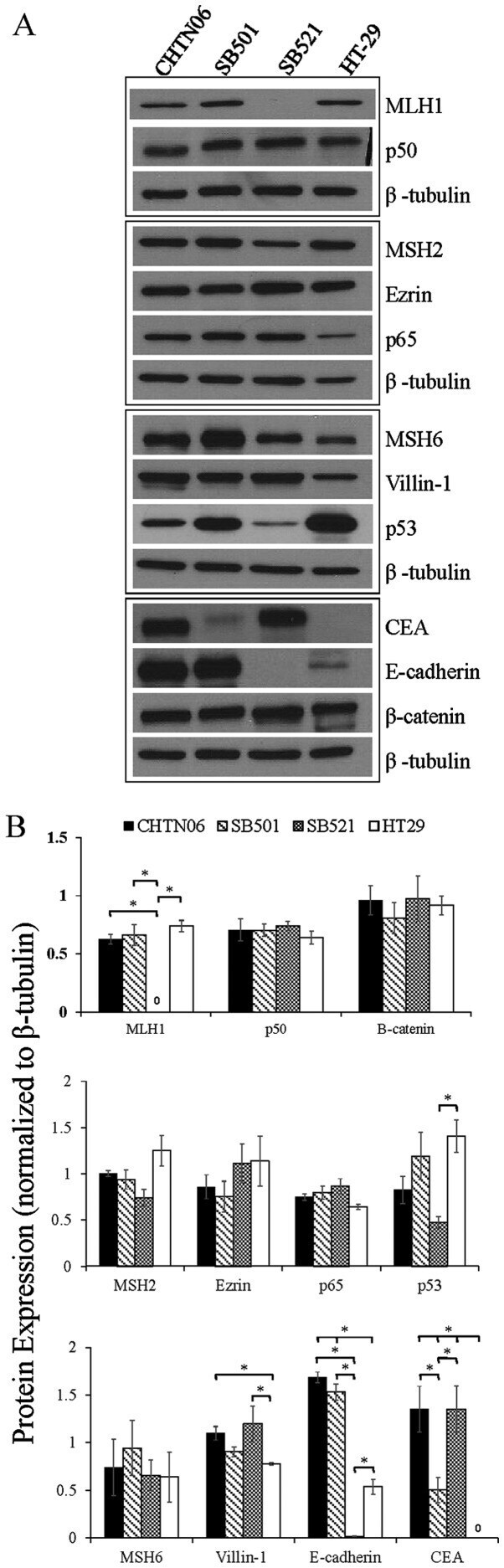
- Figure 3 The expression of proteins associated with colorectal carcinoma (CRC) tumorigenesis and metastasis was determined in the novel African American CRC lines by immunoblotting. (A) Qualitative analysis of CHTN06, SB501 and SB521 and HT-29, a Caucasian American CRC cell line, for protein expression of beta-catenin, p53, nuclear factor (NF)-kappaB (p50 and p65), villin-1, MSH2, MSH6, MLH1 and ezrin. (B) Semi-quantitative densitometry was performed by normalizing protein expression to the respective beta-tubulin loading control. Data were generated from three independent experiments. CEA, carcinoembryonic antigen.
- Submitted by
- Invitrogen Antibodies (provider)
- Main image

- Experimental details
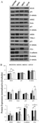
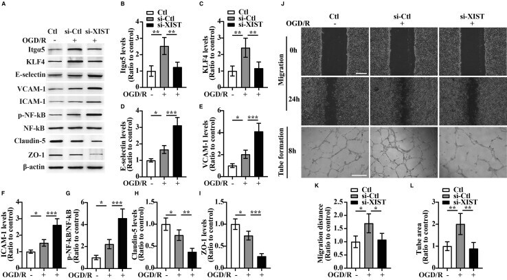
- Figure 4 The influence of silencing of lncRNA XIST on the migration and tube formation and the expression of Itgalpha5, KLF4, three CAMs, phosphorylation of (p)-NF-kappaB, and TJPs of bEnd3 cells under OGD/R conditions (A) Representative images of western blot for the expression of Itgalpha5, KLF4, three CAMs (E-selectin, VCAM-1, and ICAM-1), p-NF-kappaB, and TJPs (Claudin-5 and ZO-1) in bEnd3 cells transfected with the negative control siRNA (si-Ctl) or siRNA-lncRNA XIST (si-XIST) at 24 h restoration from OGD. The NO-OGD/R cells served as a control. (B-I) Bar graphs show the quantitative analyses of western blots as ratios of Itgalpha5/beta-actin (B), KLF4/beta-actin (C), E-selectin/beta-actin (D), VCAM-1/beta-actin (E), ICAM-1/beta-actin (F), p-NF-kappaB/total NF-kappaB (G), Claudin-5/beta-actin (H), and ZO-1/beta-actin (I) (n = 4 per experimental group). Note that the expressions of Itgalpha5, KLF4, and TJPs (Claudin-5 and ZO-1) in the si-XIST-treated bEnd3 cells were markedly reduced, but the expressions of three CAMs including E-selectin, VCAM-1, and ICAM-1 and the p-NF-kappaB were significantly elevated relative to the si-Ctl-treated group at 24 h restoration from OGD. *p < 0.05, **p < 0.01, and ***p < 0.001. (J) Representative images of cell migration and capillary tube formation of bEnd3 cells transfected with si-Ctl or si-XIST after OGD/R treatment. Scale bar, 400 mum for migration; 500 mum for tube formation. (K) Quantification of cell migration of bEnd3 cells. The
- Submitted by
- Invitrogen Antibodies (provider)
- Main image
- Experimental details
- Figure 5 Overexpression of Itgalpha5 or KLF4 reverses the effect of lncRNA XIST on the migration and tube formation and the expression of three CAMs, p-NF-kappaB, and TJPs of bEnd3 cells under OGD/R conditions (A) Representative images of western blot for the expression of Itgalpha5 in bEnd3 cells co-transfected with si-XIST and Itgalpha5 overexpression plasmid at 24 h restoration from OGD. (B) Bar graphs show the quantitative analyses of western blots as ratios of Itgalpha5/beta-actin (n = 4 per experimental group). (C) Representative images of cell migration and capillary tube formation of bEnd3 cells co-transfected with si-XIST and Itgalpha5 overexpression plasmid after OGD/R treatment. Scale bar, 400 mum for migration; 500 mum for tube formation. (D and E) Quantification of cell migration (D) and tube formation (E) of bEnd3 cells (n = 4 per experimental group). (F) Representative images of western blot for the expression of KLF4, three CAMs (E-selectin, VCAM-1, and ICAM-1), p-NF-kappaB, and TJPs (Claudin-5 and ZO-1) in bEnd3 cells co-transfected with si-XIST and KLF4 overexpression plasmid at 24 h restoration from OGD. (G-M) Bar graphs show the quantitative analyses of western blots as ratios of KLF4/beta-actin (G), E-selectin/beta-actin (H), VCAM-1/beta-actin (I), ICAM-1/beta-actin (J), p-NF-kappaB/total NF-kappaB (K), Claudin-5/beta-actin (L), and ZO-1/beta-actin (M) (n = 4 per experimental group). Note that bEnd3 cells co-transfected with si-XIST and Itgalpha5 displaye

- Submitted by
- Invitrogen Antibodies (provider)
- Main image

- Experimental details
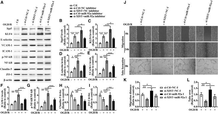
- Figure 6 lncRNA XIST alleviates a vascular endothelial inflammation response and regulates migration and tube formation of bEnd3 cells under OGD/R conditions by targeting miR-92a (A) Representative images of western blot for the expression of Itgalpha5, KLF4, E-selectin, VCAM-1, ICAM-1, p-NF-kappaB, Claudin-5, and ZO-1 in bEnd3 cells co-transfected with the si-Ctl or si-XIST and/or miR-92a inhibitor (miR-92a-I) at 24 h restoration from OGD. (B-I) Bar graphs show the quantitative analyses of western blots as ratios of Itgalpha5/beta-actin (B), KLF4/beta-actin (C), E-selectin/beta-actin (D), VCAM-1/beta-actin (E), ICAM-1/beta-actin (F), p-NF-kappaB/total NF-kappaB (G), Claudin-5/beta-actin (H), and ZO-1/beta-actin (I) (n = 4 per experimental group). Note that the expressions of Itgalpha5, KLF4, and TJPs (Claudin-5 and ZO-1) in the si-XIST + NC inhibitor (NC-I)-treated bEnd3 cells markedly decreased, but the expressions of three CAMs including E-selectin, VCAM-1, and ICAM-1 and the p-NF-kappaB were significantly elevated relative to the si-Ctl + NC-I-treated group at 24 h restoration from OGD. However, these effects were significantly rescued by inhibiting the levels of miR-92a in the bEnd3 cells. *p < 0.05, **p < 0.01, and ***p < 0.001. (J) Representative images of cell migration and capillary tube formation of bEnd3 cells transfected with si-Ctl or si-XIST and/or miR-92a-I after OGD/R treatment. Scale bar, 400 mum for migration; 500 mum for tube formation. (K and L) Quantifica
- Submitted by
- Invitrogen Antibodies (provider)
- Main image
- Experimental details
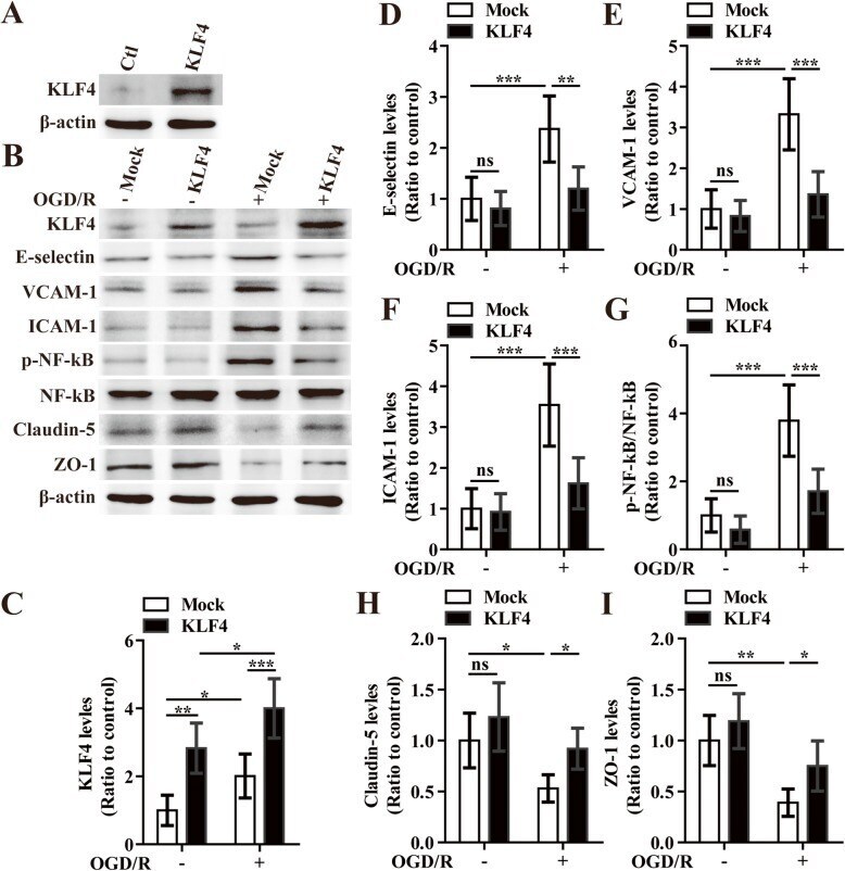
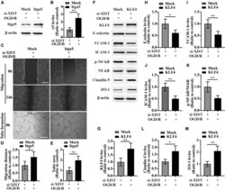
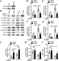
- The effect of overexpression of KLF4 on the expression of three cell adhesion molecules, phosphorylated NF-kappaB, and tight junction proteins in bEnd3 cells under OGD/R conditions. a Representative images of western blot for the expression of KLF4 in the bEnd3 cells in the control plasmid (mock) or KLF4 overexpression group. b Representative images of western blot for the expression of KLF4, E-selectin, VCAM-1, ICAM-1, p-NF-kappaB, Claudin-5, and ZO-1 in the bEnd3 cells of mock and KLF4 overexpression group at 12 h restoration of OGD or NO-OGD/R. c - i Densitometric analysis shows the ratios of KLF4/beta-actin ( c ), E-selectin/beta-actin ( d ), VCAM-1/beta-actin ( e ), ICAM-1/beta-actin ( f ), phosphorylated NF-kappaB/total NF-kappaB ( g ), Claudin-5/beta-actin ( h ), and ZO-1/beta-actin ( i ). NO-OGD/R mock-treated cells served as control. Data represent mean +- standard deviation and were analyzed by two-way ANOVA ( n = 5 per experimental group). Note that the expressions of three cell adhesion molecules including E-selectin, VCAM-1, and ICAM-1 and the phosphorylation of NF-kappaB on mock-treated bEnd3 cells were significantly induced in response to OGD/R, but the expression of Claudin-5 and ZO-1 markedly decreased compared to that of the NO-OGD/R mock-treated controls. However, these effects were significantly rescued by overexpressing the levels of KLF4 in the bEnd3 cells. * P < 0.05, ** P < 0.01, *** P < 0.001; ns, not significant