Explore
 Validate
 Learn
Learn710450
antibody from Invitrogen Antibodies
Targeting: NFKB1
KBF1, NF-kappaB, NF-kB1, NFkappaB, NFKB-p50, p105, p50
 Western blot
 Immunocytochemistry
Antibody data
- Antibody Data
- Antigen structure
- References [1]
- Comments [0]
- Validations
- Immunocytochemistry [4]
- Immunohistochemistry [3]
- Chromatin Immunoprecipitation [2]
- Other assay [2]
Submit
Validation data
Reference
Comment
Report error
- Product number
- 710450 - Provider product page

- Provider
- Invitrogen Antibodies
- Product name
- NFkB p50 Recombinant Superclonal™ Antibody (24HCLC)
- Antibody type
- Other
- Antigen
- Synthetic peptide
- Description
- Recombinant rabbit Superclonal™ antibodies are unique offerings from Thermo Fisher Scientific. They are comprised of a selection of multiple different recombinant monoclonal antibodies, providing the best of both worlds - the sensitivity of polyclonal antibodies with the specificity of monoclonal antibodies - all delivered with the consistency only found in a recombinant antibody. While functionally the same as a polyclonal antibody - recognizing multiple epitope sites on the target and producing higher detection sensitivity for low abundance targets - a recombinant rabbit Superclonal™ antibody has a known mixture of light and heavy chains. The exact population can be produced in every lot, circumventing the biological variability typically associated with polyclonal antibody production. Note: Formerly called “Recombinant polyclonal antibody”, this product is now rebranded as “Recombinant Superclonal™ antibody”. The physical product and the performance remain unchanged.
- Reactivity
- Human, Mouse
- Host
- Rabbit
- Isotype
- IgG
- Antibody clone number
- 24HCLC
- Vial size
- 100 μg
- Concentration
- 0.5 mg/mL
- Storage
- Store at 4°C short term. For long term storage, store at -20°C, avoiding freeze/thaw cycles.
Submitted references Regulatory role of DEPTOR‑mediated cellular autophagy and mitochondrial reactive oxygen species in angiogenesis in multiple myeloma.
Wang J, Chen J, Qiu D, Zeng Z
International journal of molecular medicine 2021 Feb;47(2):643-658
International journal of molecular medicine 2021 Feb;47(2):643-658
No comments: Submit comment
Supportive validation
- Submitted by
- Invitrogen Antibodies (provider)
- Main image
- Experimental details
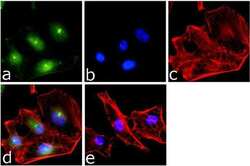
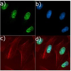
- Immunofluorescence analysis of NFkB p50 was done on 70% confluent log phase HeLa cells. The cells were fixed with 4% paraformaldehyde for 15 minutes, permeabilized with 0.25% Triton™ X-100 for 10 minutes, and blocked with 5% BSA for 1 hour at room temperature. The cells were labeled with NFkB p50 (24HCLC), Recombinant Rabbit Polyclonal Antibody (Product # 710450) at 1 µg/mL in 1% BSA and incubated for 3 hours at room temperature and then labeled with Goat anti-Rabbit IgG (H+L) Superclonal™ Secondary Antibody, Alexa Fluor® 488 conjugate (Product # A27034) at a dilution of 1:2000 for 45 minutes at room temperature (Panel a: green). Nuclei (Panel b: blue) were stained with SlowFade® Gold Antifade Mountant with DAPI (Product # S36938). F-actin (Panel c: red) was stained with Alexa Fluor® 555 Rhodamine Phalloidin (Product # R415, 1:300). Panel d is a merged image showing Nuclear localization. Panel e is a no primary antibody control. The images were captured at 60X magnification.

- Submitted by
- Invitrogen Antibodies (provider)
- Main image

- Experimental details
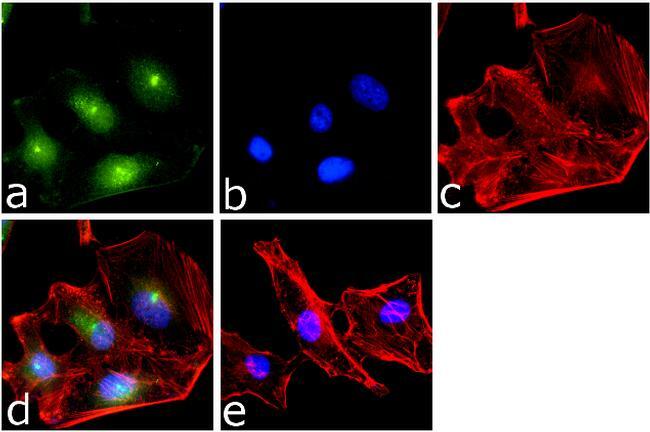
- Immunofluorescent analysis of NFkB p50 in HeLa cells using a NFkB p50 Recombinant Rabbit Polyclonal Antibody (Product # 710450) followed by detection using an Alexa Fluor 488-conjugated Goat anti-Rabbit secondary antibody (green) (Image A). Nuclei were stained using DAPI (Image B) and actin stained with Alexa Fluor 594 phalloidin (red) (image C). Image D is a composite image showing nuclear localization of p50.
- Submitted by
- Invitrogen Antibodies (provider)
- Main image

- Experimental details
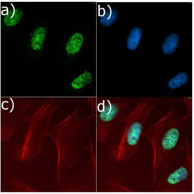
- Immunofluorescence analysis of NFkB p50 was done on 70% confluent log phase HeLa cells. The cells were fixed with 4% paraformaldehyde for 15 minutes, permeabilized with 0.25% Triton™ X-100 for 10 minutes, and blocked with 5% BSA for 1 hour at room temperature. The cells were labeled with NFkB p50 (24HCLC), Recombinant Rabbit Superclonal™ Antibody (Product # 710450) at 1 µg/mL in 1% BSA and incubated for 3 hours at room temperature and then labeled with Goat anti-Rabbit IgG (Heavy Chain) Superclonal™ Secondary Antibody, Alexa Fluor® 488 conjugate (Product # A27034) at a dilution of 1:2000 for 45 minutes at room temperature (Panel a: green). Nuclei (Panel b: blue) were stained with SlowFade® Gold Antifade Mountant with DAPI (Product # S36938). F-actin (Panel c: red) was stained with Alexa Fluor® 555 Rhodamine Phalloidin (Product # R415, 1:300). Panel d is a merged image showing Nuclear localization. Panel e is a no primary antibody control. The images were captured at 60X magnification.
- Submitted by
- Invitrogen Antibodies (provider)
- Main image
- Experimental details
- Immunofluorescent analysis of NFkB p50 in HeLa cells using a NFkB p50 Recombinant Rabbit Superclonal™ Antibody (Product # 710450) followed by detection using an Alexa Fluor 488-conjugated Goat anti-Rabbit secondary antibody (green) (Image A). Nuclei were stained using DAPI (Image B) and actin stained with Alexa Fluor 594 phalloidin (red) (image C). Image D is a composite image showing nuclear localization of p50.

Supportive validation
- Submitted by
- Invitrogen Antibodies (provider)
- Main image
- Experimental details
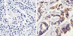
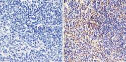
- Immunohistochemistry analysis of NFkB p50 showing staining in the cytoplasm of paraffin-embedded human breast carcinoma (right) compared to a negative control without primary antibody (left). To expose target proteins, antigen retrieval was performed using 10 mM sodium citrate (pH 6.0), microwaved for 8-15 min. Following antigen retrieval, tissues were blocked in 3% H2O2-methanol for 15 min at room temperature, washed with ddH2O and PBS, and then probed with NFkB p50 Recombinant Rabbit Superclonal™ Antibody (Product # 710450) diluted in 3% BSA-PBS at a dilution of 1:100 overnight at 4°C in a humidified chamber. Tissues were washed extensively in PBST and detection was performed using a HRP-conjugated secondary antibody followed by colorimetric detection using a DAB kit. Tissues were counterstained with hematoxylin and dehydrated with ethanol and xylene to prep for mounting.
- Submitted by
- Invitrogen Antibodies (provider)
- Main image
- Experimental details
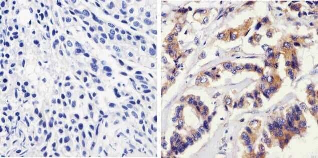
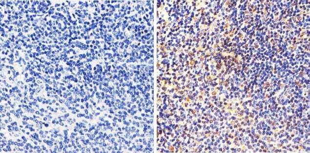
- Immunohistochemistry analysis of NFkB p50 showing staining in the cytoplasm and nucleus of paraffin-embedded human tonsil tissue (right) compared to a negative control without primary antibody (left). To expose target proteins, antigen retrieval was performed using 10 mM sodium citrate (pH 6.0), microwaved for 8-15 min. Following antigen retrieval, tissues were blocked in 3% H2O2-methanol for 15 min at room temperature, washed with ddH2O and PBS, and then probed with NFkB p50 Recombinant Rabbit Superclonal™ Antibody (Product # 710450) diluted in 3% BSA-PBS at a dilution of 1:100 overnight at 4°C in a humidified chamber. Tissues were washed extensively in PBST and detection was performed using a HRP-conjugated secondary antibody followed by colorimetric detection using a DAB kit. Tissues were counterstained with hematoxylin and dehydrated with ethanol and xylene to prep for mounting.
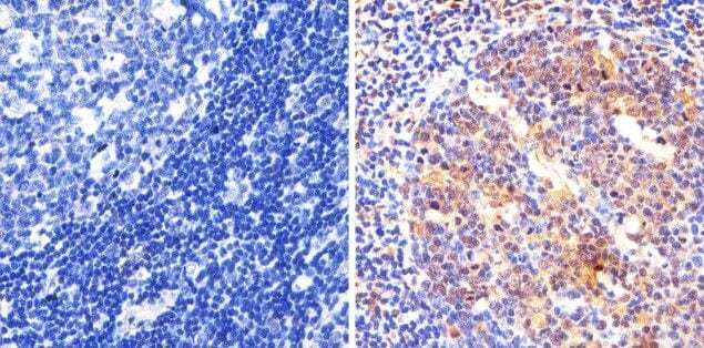
- Submitted by
- Invitrogen Antibodies (provider)
- Main image
- Experimental details
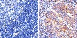
- Immunohistochemistry analysis of NFkB p50 showing staining in the cytoplasm and nucleus of paraffin-embedded mouse spleen tissue (right) compared to a negative control without primary antibody (left). To expose target proteins, antigen retrieval was performed using 10 mM sodium citrate (pH 6.0), microwaved for 8-15 min. Following antigen retrieval, tissues were blocked in 3% H2O2-methanol for 15 min at room temperature, washed with ddH2O and PBS, and then probed with a NFkB p50 Recombinant Rabbit Superclonal™ Antibody (Product # 710450) diluted in 3% BSA-PBS at a dilution of 1:20 overnight at 4°C in a humidified chamber. Tissues were washed extensively in PBST and detection was performed using a HRP-conjugated secondary antibody followed by colorimetric detection using a DAB kit. Tissues were counterstained with hematoxylin and dehydrated with ethanol and xylene to prep for mounting.
Supportive validation
- Submitted by
- Invitrogen Antibodies (provider)
- Main image
- Experimental details
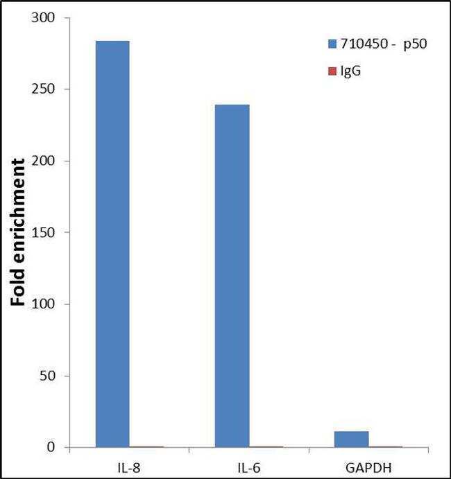
- Enrichment of endogenous NFkB p50 protein at specific gene loci using Anti-NFkB p50 Recombinant Rabbit Polyclonal Antibody: Chromatin Immunoprecipitation (ChIP) was performed using Anti-NFkB p50 Recombinant Rabbit Polyclonal Antibody (Product # 710450, 5 µg) on sheared chromatin from 2 million HeLa cells treated with 50 ng/mL of TNFalpha for 45 minutes using the "MAGnify ChIP system" kit (Product # 49-2024). Normal Rabbit IgG was used as a negative IP control. The purified DNA was analyzed by 7500 Fast qPCR system (Product # 4351106) with optimized PCR primer pairs for the promoter of active IL-8, IL-6 gene, used as positive control target, and the GAPDH, used as negative control target. Data is presented as fold enrichment of the antibody signal versus the negative control IgG using the comparative CT method.
- Submitted by
- Invitrogen Antibodies (provider)
- Main image

- Experimental details
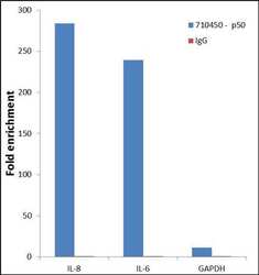
- Enrichment of endogenous NFkB p50 protein at specific gene loci using Anti-NFkB p50 Recombinant Rabbit Superclonal™ Antibody: Chromatin Immunoprecipitation (ChIP) was performed using Anti-NFkB p50 Recombinant Rabbit Superclonal™ Antibody (Product # 710450, 5 µg) on sheared chromatin from 2 million HeLa cells treated with 50 ng/mL of TNFalpha for 45 minutes using the "MAGnify ChIP system" kit (Product # 49-2024). Normal Rabbit IgG was used as a negative IP control. The purified DNA was analyzed by 7500 Fast qPCR system (Product # 4351106) with optimized PCR primer pairs for the promoter of active IL-8, IL-6 gene, used as positive control target, and the GAPDH, used as negative control target. Data is presented as fold enrichment of the antibody signal versus the negative control IgG using the comparative CT method.

Supportive validation
- Submitted by
- Invitrogen Antibodies (provider)
- Main image
- Experimental details
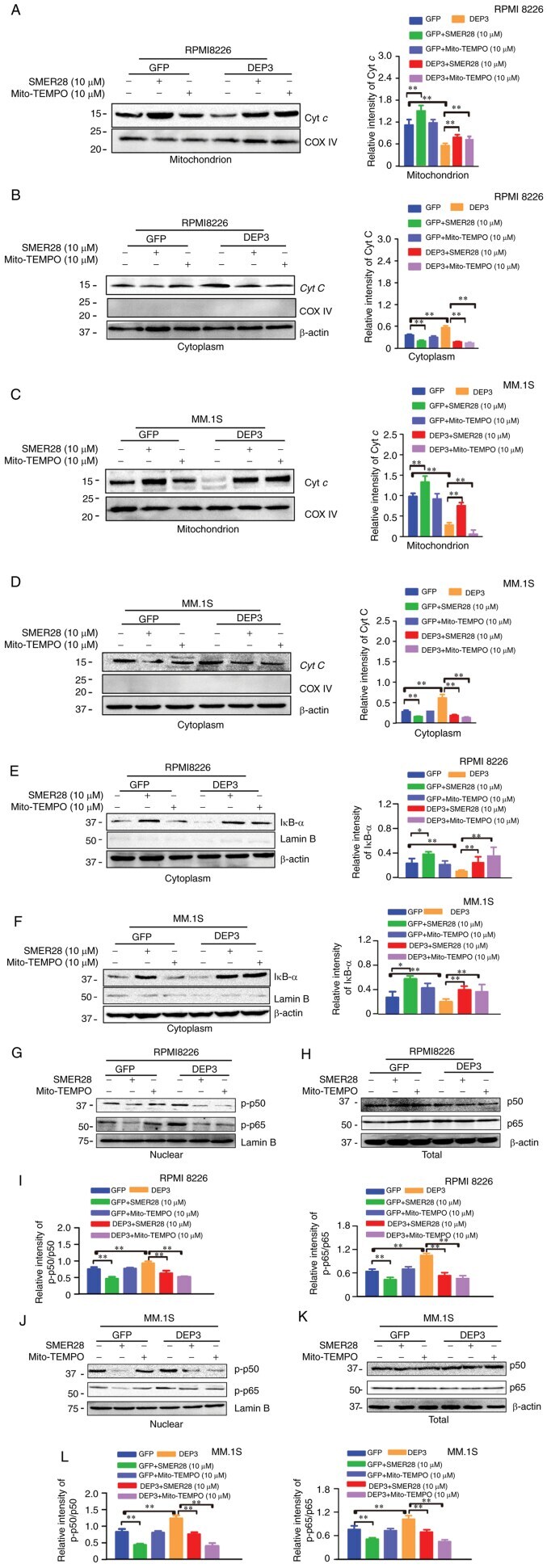
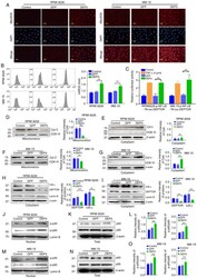
- Figure 4 Mitochondrial damage signals in DEPTOR-inhibited RPMI8226 and MM.1S cells. RPMI8226 and MM.1s cells were infected with lentiviral GFP and DEP3 for 5 days. (A and B) The mtROS levels of the RPMI8226 and MM.1S cells in the indicated groups stained with the MitoSOX probe was evaluated using a fluorescence microscope (scale bar, 10 u m) and flow cytometric analysis, respectively. (C) The NF-kappaB activity of RPMI8226 or MM.1S cells in the indicated groups was detected by luciferase assays. Cells were co-transfected with pNF-kappaB-TA-luc with Renilla luciferase reporter (as an internal control) for 24 h and treated with GFP or DEP3 for 48 h. (D-G) The mitochondrial and cytoplasmic fractions of the cultured cells were separated using the Mitochondria/Cytosol Fractionation kit. Protein expression of Cyt c in the (D) mitochondria and (E) cytoplasm of the indicated groups for RPMI8226 cells was examined by western blot analysis. Primary antibodies with COXIV and beta-actin was used as the mitochondrial and cytosolic loading control, respectively. Protein expression of Cyt c in the (F) mitochondria and (G) cytoplasm of the indicated groups for MM.1S cells was examined by western blot analysis. Primary antibodies with COXIV and beta-actin was used as the mitochondrial and cytosolic loading control, respectively. Protein expression levels of DEPTOR and IkappaB-alpha in the cytoplasm of the indicated groups for (H) RPMI8226 and (I) MM.1S cells were examined by western blot anal

- Submitted by
- Invitrogen Antibodies (provider)
- Main image
- Experimental details
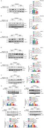
- Figure 6 Autophagy inducer and mitochondrial-specific antioxidants reversed the effects of DEPTOR on the angiogenesis of MM cells. RPMI 8226 and MM.1S cells were transfected with lentivirus DEP3 for 5 days and exposed to SMER28 (10 u M) or Mito-TEMPO (10 u M) for 24 h. (A-D) The mitochondrial and cytoplasmic fractions of the cultured cells were separated using the Mitochondria/Cytosol Fractionation kit. Protein expression of Cyt c in the (A) mitochondria and (B) cytoplasm of the indicated groups for RPMI8226 cells was examined by western blot analysis, respectively. Primary antibodies with COXIV and beta-actin was used as the mitochondrial and cytosolic loading control, respectively. Protein expression of Cyt c in the (C) mitochondria and (D) cytoplasm of the indicated groups for MM.1S was examined by western blot analysis, respectively. Primary antibodies with COXIV and beta-actin was used as the mitochondrial and cytosolic loading control, respectively. Protein expression levels of DEPTOR and IkappaB-alpha in the cytoplasm of the indicated groups for (E) RPMI8226 and (F) MM.1S cells were examined by western blot analysis, respectively. (G-L) The nuclear and cytoplasmic protein fractions of the cultured cells were isolated using the NE-PER(tm) Nuclear and Cytoplasmic Extraction Reagents. Protein expressions of (G) p-p50 and p-p65 in the nuclear, (H) p50 and p65 in the whole cell lysates of the indicated groups for RPMI8226 cells was examined by western blot analysis, respect