Explore
 Validate
 Learn
Learn710460
antibody from Invitrogen Antibodies
Targeting: NFKB1
KBF1, NF-kappaB, NF-kB1, NFkappaB, NFKB-p50, p105, p50
 Western blot
 ELISA
ELISAAntibody data
- Antibody Data
- Antigen structure
- References [1]
- Comments [0]
- Validations
- Western blot [2]
- Chromatin Immunoprecipitation [1]
- Other assay [1]
Submit
Validation data
Reference
Comment
Report error
- Product number
- 710460 - Provider product page

- Provider
- Invitrogen Antibodies
- Product name
- Phospho-NFkB p50 (Ser337) Recombinant Polyclonal Antibody (2HCLC)
- Antibody type
- Polyclonal
- Antigen
- Synthetic peptide
- Description
- Recombinant rabbit polyclonal antibodies are unique offerings from Thermo Fisher Scientific. They are comprised of a selection of multiple different recombinant monoclonal antibodies, providing the best of both worlds - the sensitivity of polyclonal antibodies with the specificity of monoclonal antibodies - all delivered with the consistency only found in a recombinant antibody. While functionally the same as a polyclonal antibody - recognizing multiple epitope sites on the target and producing higher detection sensitivity for low abundance targets - a recombinant rabbit polyclonal antibody has a known mixture of light and heavy chains. The exact population can be produced in every lot, circumventing the biological variability typically associated with polyclonal antibody production.
- Reactivity
- Human
- Host
- Rabbit
- Isotype
- IgG
- Antibody clone number
- 2HCLC
- Vial size
- 100 µg
- Concentration
- 0.5 mg/mL
- Storage
- Store at 4°C short term. For long term storage, store at -20°C, avoiding freeze/thaw cycles.
Submitted references The role of NFκB in spheroid formation of human breast cancer cells cultured on the Random Positioning Machine.
Kopp S, Sahana J, Islam T, Petersen AG, Bauer J, Corydon TJ, Schulz H, Saar K, Huebner N, Slumstrup L, Riwaldt S, Wehland M, Infanger M, Luetzenberg R, Grimm D
Scientific reports 2018 Jan 17;8(1):921
Scientific reports 2018 Jan 17;8(1):921
No comments: Submit comment
Supportive validation
- Submitted by
- Invitrogen Antibodies (provider)
- Main image

- Experimental details
- Western blot analysis was performed on whole cell extracts (30 µg lysate) of Jurkat (Lane 1), Jurkat treated with TNF alpha (Lane 2). The blots were probed with Recombinant Rabbit Polyclonal Anti-NFkB p50 (pS337) Antibody (Product # 710460, 1-2 µg/mL) and detected by chemiluminescence using Goat anti-Rabbit IgG (H+L) Superclonal™ Secondary Antibody, HRP conjugate (Product # A27036, 0.4 µg/mL, 1:2500 dilution). A 55 kDa band corresponding to NFkB p50 (pS337)was observed across cell lines tested. Known quantity of protein samples were electrophoresed using Novex® NuPAGE® 4-12 % Bis-Tris gel (Product # NP0321BOX), XCell SureLock™ Electrophoresis System (Product # EI0002) and Novex® Sharp Pre-Stained Protein Standard (Product # LC5800). Resolved proteins were then transferred onto a nitrocellulose membrane with iBlot® 2 Dry Blotting System (Product # IB21001). The membrane was probed with the relevant primary and secondary Antibody following blocking with 5 % skimmed milk. Chemiluminescent detection was performed using Pierce™ ECL Western blotting Substrate (Product # 32106).

- Submitted by
- Invitrogen Antibodies (provider)
- Main image

- Experimental details
- Western blot analysis of Phospho-NFkB p50 pSer337 in whole cell extracts from TNFa treated Jurkat (20 ng/mL, 15 min) using a Phospho-NFkB p50 pSer337 Recombinant Rabbit Polyclonal Antibody (Product # 710460) at a dilution of 1 µg/mL. Detection was performed using an HRP-conjugated Goat anti-Rabbit secondary antibody followed by chemiluminescence (ECL). Results show a band at ~50 kDa.

Supportive validation
- Submitted by
- Invitrogen Antibodies (provider)
- Main image

- Experimental details
- Enrichment of endogenous NFkB p50 pSer337 protein at specific gene loci using Anti-Phospho-NFkB p50 pSer337 Recombinant Rabbit Polyclonal Antibody: Chromatin Immunoprecipitation (ChIP) was performed using Anti-Phospho-NFkB p50 pSer337 Recombinant Rabbit Polyclonal Antibody (Product # 710460, 5 µg) on sheared chromatin from 2 million HeLa cells treated with 50 ng/mL of TNFalpha for 45 minutes using the MAGnify ChIP system kit (Product # 49-2024). Normal Rabbit IgG was used as a negative IP control. The purified DNA was analyzed by 7500 Fast qPCR system (Product # 4351106) with optimized PCR primer pairs for the promoter of active IL-8, IkB gene, used as positive control target, and the SAT2, used as negative control target. Data is presented as fold enrichment of the antibody signal versus the negative control IgG using the comparative CT method.

Supportive validation
- Submitted by
- Invitrogen Antibodies (provider)
- Main image

- Experimental details
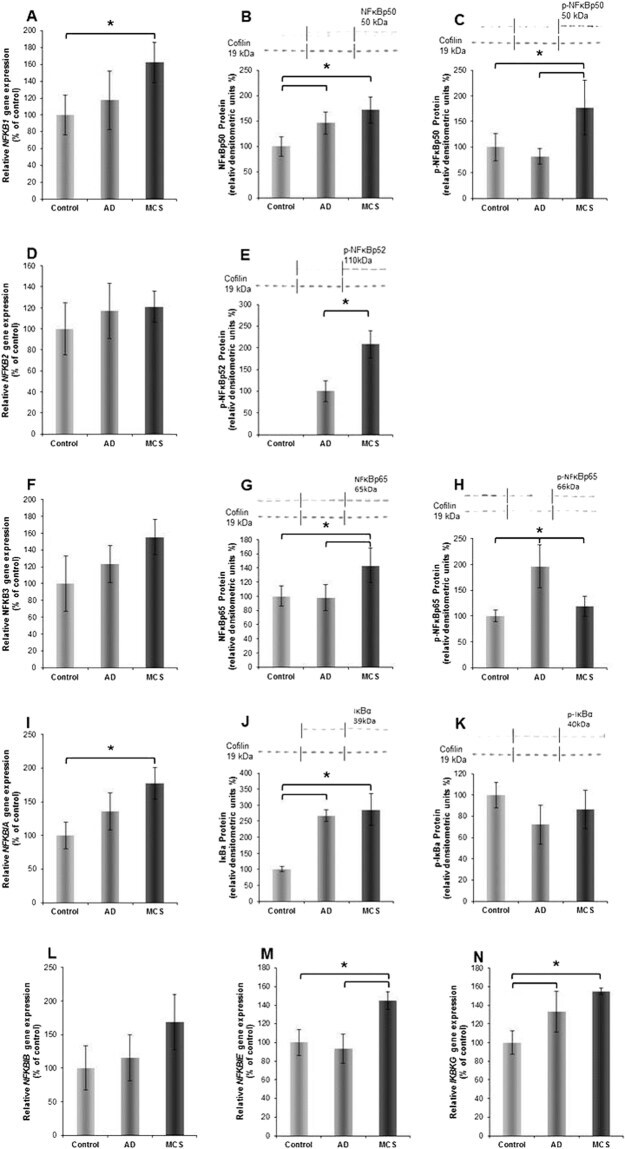
- Figure 2 ( A ) NFKB1 gene expression; ( B ) NFkBp50 Western blot analysis; ( C ) phosphorylated (p)-NFkappaBp50 Western blot analysis; ( D ) NFKB2 gene expression; ( E ) p-NFkappaBp52 Western blot analysis; ( F ) NFKB3 gene expression; ( G ) NFkappaBp65 Western blot analysis; ( H ) p-NFkBp65 Western blot analysis; ( I ) NFKBIA gene expression; ( J ) IkappaBalpha Western blot analysis; ( K ) p- IkappaBbeta Western blot analysis; ( L ) NFKBIB gene expression; ( M ) NFKBIE gene expression; ( N ) IKBKG gene expression. The position (arrow) and molecular size (in kD) of the investigated proteins are indicated on each of the Western blot membrane images. Cofilin 1 was used as loading control. The vertical lines indicate group separation giving n = 5 per group.