Explore
 Validate
 Learn
Learn Western blot
 Immunocytochemistry
Antibody data
- Antibody Data
- Antigen structure
- References [1]
- Comments [0]
- Validations
- Immunocytochemistry [5]
- Immunohistochemistry [2]
- Other assay [1]
Submit
Validation data
Reference
Comment
Report error
- Product number
- PA5-27592 - Provider product page

- Provider
- Invitrogen Antibodies
- Product name
- HAX1 Polyclonal Antibody
- Antibody type
- Polyclonal
- Antigen
- Recombinant full-length protein
- Description
- Recommended positive controls: Molt-4, Raji, 293T, A431 , HeLa, HepG2. Predicted reactivity: Mouse (80%), Rhesus Monkey (97%), Bovine (83%). Store product as a concentrated solution. Centrifuge briefly prior to opening the vial.
- Reactivity
- Human, Mouse, Rat
- Host
- Rabbit
- Isotype
- IgG
- Vial size
- 100 μL
- Concentration
- 2.87 mg/mL
- Storage
- Store at 4°C short term. For long term storage, store at -20°C, avoiding freeze/thaw cycles.
Submitted references The interactome of multifunctional HAX1 protein suggests its role in the regulation of energy metabolism, de-aggregation, cytoskeleton organization and RNA-processing.
Wakula M, Balcerak A, Rubel T, Chmielarczyk M, Konopinski R, Lyczek F, Grzybowska EA
Bioscience reports 2020 Nov 27;40(11)
Bioscience reports 2020 Nov 27;40(11)
No comments: Submit comment
Supportive validation
- Submitted by
- Invitrogen Antibodies (provider)
- Main image
- Experimental details
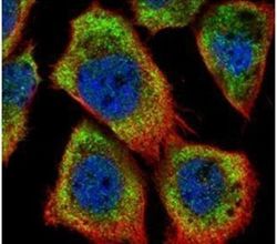
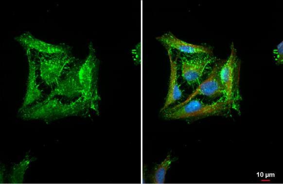
- Immunofluorescent analysis of HAX1 in methanol-fixed A431 cells using a HAX1 polyclonal antibody (Product # PA5-27592) (Green) at a 1:500 dilution. Alpha-tubulin filaments were labeled with Product # PA5-29281 (Red) at a 1:2000.

- Submitted by
- Invitrogen Antibodies (provider)
- Main image
- Experimental details
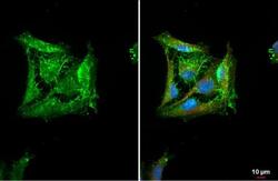
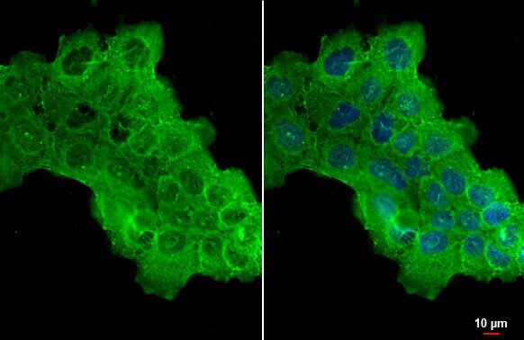
- HAX1 Polyclonal Antibody detects HAX1 protein at cytoplasm by immunofluorescent analysis. Sample: A431 cells were fixed in ice-cold MeOH for 5 min. Green: HAX1 stained by HAX1 Polyclonal Antibody (Product # PA5-27592) diluted at 1:500.

- Submitted by
- Invitrogen Antibodies (provider)
- Main image
- Experimental details
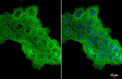
- HAX1 Polyclonal Antibody detects HAX1 protein at cell membrane by immunofluorescent analysis. Sample: HeLa cells were fixed in ice-cold MeOH for 5 min. Green: HAX1 stained by HAX1 Polyclonal Antibody (Product # PA5-27592) diluted at 1:500. Red: alpha Tubulin, a cytoskeleton marker, stained by alpha Tubulin Polyclonal Antibody [GT114] (Product # MA5-31466) diluted at 1:1,000. Blue: Fluoroshield with DAPI .

- Submitted by
- Invitrogen Antibodies (provider)
- Main image

- Experimental details
- HAX1 Polyclonal Antibody detects HAX1 protein at cytoplasm by immunofluorescent analysis. Sample: A431 cells were fixed in ice-cold MeOH for 5 min. Green: HAX1 stained by HAX1 Polyclonal Antibody (Product # PA5-27592) diluted at 1:500.
- Submitted by
- Invitrogen Antibodies (provider)
- Main image

- Experimental details
- HAX1 Polyclonal Antibody detects HAX1 protein at cell membrane by immunofluorescent analysis. Sample: HeLa cells were fixed in ice-cold MeOH for 5 min. Green: HAX1 stained by HAX1 Polyclonal Antibody (Product # PA5-27592) diluted at 1:500. Red: alpha Tubulin, a cytoskeleton marker, stained by alpha Tubulin Polyclonal Antibody [GT114] (Product # MA5-31466) diluted at 1:1,000. Blue: Fluoroshield with DAPI .
Supportive validation
- Submitted by
- Invitrogen Antibodies (provider)
- Main image

- Experimental details
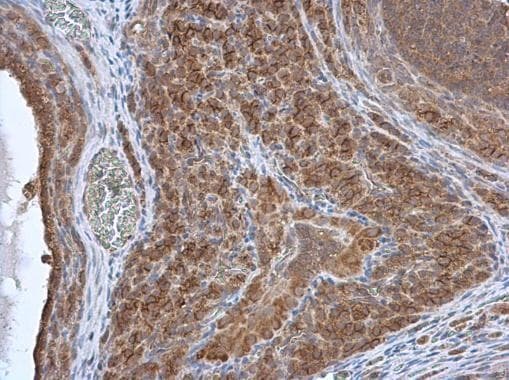
- Immunohistochemistry (Paraffin) analysis of HAX1 was performed in paraffin-embedded rat ovary tissue using HAX1 Polyclonal Antibody (Product # PA5-27592) at a dilution of 1:500.
- Submitted by
- Invitrogen Antibodies (provider)
- Main image
- Experimental details
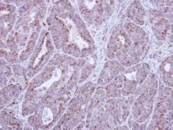
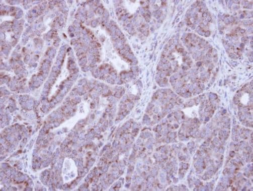
- Immunohistochemical analysis of paraffin-embedded NCIN87 Xenograft , using HAX1 (Product # PA5-27592) antibody at 1:100 dilution. Antigen Retrieval: Citrate buffer, pH 6.0, 15 min.
Supportive validation
- Submitted by
- Invitrogen Antibodies (provider)
- Main image
- Experimental details
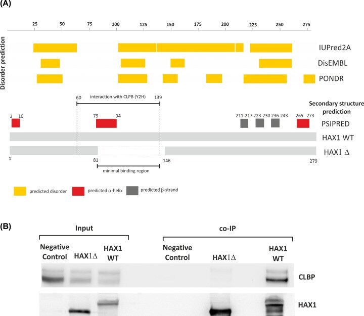
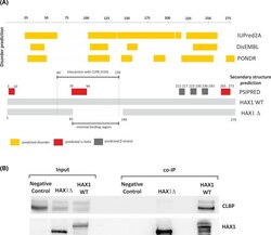
- Figure 5 Mapping of the HAX1-CLPB interaction ( A ) Prediction of secondary structure elements and structural disorder in HAX1 in relation to CLPB binding region. The region predicted as interacting with CLPB according to Y2H, the region of the deletion which precludes CLPB binding (81-146 aa) and the minimal binding region are marked. ( B ) Co-immunoprecipitation with the whole HAX1 and the 81-146 aa deletion, showing minimal interaction for the deletion.