Explore
 Validate
 Learn
Learn Western blot
 Immunohistochemistry
Antibody data
- Antibody Data
- Antigen structure
- References [0]
- Comments [0]
- Validations
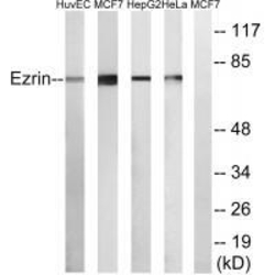
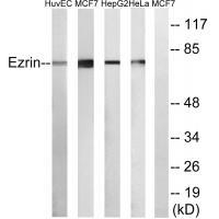
- Western blot [1]
Submit
Validation data
Reference
Comment
Report error
- Product number
- A01750 - Provider product page

- Provider
- Boster Biological Technology
- Product name
- Anti-Ezrin Antibody
- Antibody type
- Polyclonal
- Description
- Polyclonal antibody for Ezrin/EZR detection. Host: Rabbit.Size: 100μl. Tested applications: IHC. Reactive species: Human. Ezrin/EZR information: Molecular Weight: 69413 MW; Subcellular Localization: Apical cell membrane ; Peripheral membrane protein ; Cytoplasmic side . Cell projection . Cell projection, microvillus membrane ; Peripheral membrane protein ; Cytoplasmic side . Cell projection, ruffle membrane ; Peripheral membrane protein ; Cytoplasmic side . Cytoplasm, cell cortex . Cytoplasm, cytoskeleton . Localization to the apical membrane of parietal cells depends on the interaction with MPP5. Localizes to cell extensions and peripheral processes of astrocytes (By similarity). Microvillar peripheral membrane protein (cytoplasmic side); Tissue Specificity: Expressed in cerebral cortex, basal ganglia, hippocampus, hypophysis, and optic nerve. Weakly expressed in brain stem and diencephalon. Stronger expression was detected in gray matter of frontal lobe compared to white matter (at protein level). Component of the microvilli of intestinal epithelial cells. Preferentially expressed in astrocytes of hippocampus, frontal cortex, thalamus, parahippocampal cortex, amygdala, insula, and corpus callosum. Not detected in neurons in most tissues studied.
- Reactivity
- Human, Mouse, Rat
- Host
- Rabbit
- Vial size
- 100μl
- Concentration
- 0.5-1mg/ml, actual concentration vary by lot. Use suggested dilution ratio to decide dilution procedure.
- Storage
- Store at -20°C for one year, at 4°C for one month. We suggest the antibody be aliquoted into small vials and stored frozen at -20°C upon receiving. Avoid repeated freezing and thawing.
No comments: Submit comment
Supportive validation
- Submitted by
- Boster Biological Technology (provider)
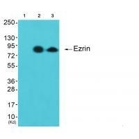
- Main image
- Experimental details
- Western blot analysis of extracts from cos-7 cells (Lane 2) and 3T3 cells (Lane 3), using Ezrin antiobdy A01750. The lane on the left is treated with synthesized peptide.
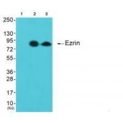
- Additional image