 Explore
 Validate
 Learn
Learn Western blot
 Immunoprecipitation
Antibody data
- Antibody Data
- Antigen structure
- References [2]
- Comments [0]
- Validations
- Western blot [4]
- Immunohistochemistry [1]
- Other assay [1]
Submit
Validation data
Reference
Comment
Report error
- Product number
- PA5-17518 - Provider product page

- Provider
- Invitrogen Antibodies
- Product name
- Ezrin Polyclonal Antibody
- Antibody type
- Polyclonal
- Antigen
- Synthetic peptide
- Description
- It is not recommended to aliquot this antibody. This antibody is not cross-reactive with ezrin homologues such as radixin and moesin.
- Reactivity
- Human, Mouse, Rat, Bovine
- Host
- Rabbit
- Isotype
- IgG
- Vial size
- 100 µL
- Concentration
- 12 µg/mL
- Storage
- -20°C
Submitted references An in vitro intestinal platform with a self-sustaining oxygen gradient to study the human gut/microbiome interface.
Formation of Human Colonic Crypt Array by Application of Chemical Gradients Across a Shaped Epithelial Monolayer.
Kim R, Attayek PJ, Wang Y, Furtado KL, Tamayo R, Sims CE, Allbritton NL
Biofabrication 2019 Nov 6;12(1):015006
Biofabrication 2019 Nov 6;12(1):015006
Formation of Human Colonic Crypt Array by Application of Chemical Gradients Across a Shaped Epithelial Monolayer.
Wang Y, Kim R, Gunasekara DB, Reed MI, DiSalvo M, Nguyen DL, Bultman SJ, Sims CE, Magness ST, Allbritton NL
Cellular and molecular gastroenterology and hepatology 2018;5(2):113-130
Cellular and molecular gastroenterology and hepatology 2018;5(2):113-130
No comments: Submit comment
Supportive validation
- Submitted by
- Invitrogen Antibodies (provider)
- Main image

- Experimental details
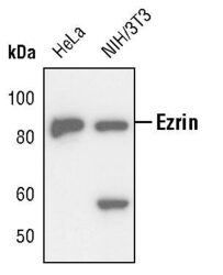
- Western blot analysis of Ezrin in extracts from HeLa and NIH/3T3 cells using Ezrin polyclonal antibody (Product # PA5-17518).

- Submitted by
- Invitrogen Antibodies (provider)
- Main image
- Experimental details
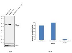
- CRISPR-Cas9 mediated genome editing ofEzrin (as confirmed by next generation sequencing) was achieved by using LentiArray™ Lentiviral sgRNA (Product # A32042, AssayID CRISPR864181_LV) and LentiArray Cas9 Lentivirus (Product # A32064). Fig (a) Western blot analysis of Ezrin was performed by loading 30 µg of HeLa Wild Type (Lane 1), HeLa Cas9 (Lane 2) and HeLa Cas9 cells transduced with Ezrin Lentiviral sgRNA (Lane 3) whole cell extracts. The samples were electrophoresed using NuPAGE™ Novex™ 4-12% Bis-Tris Protein Gel (Product # NP0322BOX). Resolved proteins were then transferred onto a nitrocellulose membrane (Product # IB23001) by iBlot® 2 Dry Blotting System (Product # IB21001). The blot was probed with Anti-Ezrin Polyclonal Antibody (Product # PA5-17518) using 1:1,000 dilution and Goat anti-Rabbit IgG (H+L) Superclonal™ Recombinant Secondary Antibody, HRP (Product # A27036 1:5,000 dilution).Chemiluminescent detection was performed using Novex® ECL Chemiluminescent Substrate Reagent Kit (Product # WP20005). A reduced signal in sgRNA transduced cells using the LentiArray™ CRISPR product line confirms that antibody is specific toEzrin (Fig (b)).

- Submitted by
- Invitrogen Antibodies (provider)
- Main image
- Experimental details
- Western blot analysis of Ezrin in extracts from HeLa and NIH/3T3 cells using Ezrin polyclonal antibody (Product # PA5-17518).

- Submitted by
- Invitrogen Antibodies (provider)
- Main image

- Experimental details
- Western blot was performed using Anti-Ezrin Polyclonal Antibody(Product # PA5-17518) and a ~70kDa band corresponding to Ezrin was observed across cell lines tested . Membrane enriched extracts (30 µg lysate) of HeLa (Lane 1), HCT 116 (Lane 2), A-431 (Lane 3), HaCaT (Lane 4), NTERA-2 cl.D1 (Lane 5), HEK-293 (Lane 6) were electrophoresed using NuPAGE™ 4-12% Bis-Tris Protein Gel (Product # NP0322BOX). Resolved proteins were then transferred onto a Nitrocellulose membrane (Product # LC2002) by iBlot® 2 Dry Blotting System (Product # IB21001). The blot was probed with the primary antibody (1:1000 dilution) and detected by chemiluminescence with Goat anti-Rabbit IgG (H+L) Superclonal™ Recombinant Secondary Antibody, HRP (Product # A27036, 1:4000) using the iBright FL 1000 (Product # A32752). Chemiluminescent detection was performed using Novex® ECL Chemiluminescent Substrate Reagent Kit (Product # WP20005).

Supportive validation
- Submitted by
- Invitrogen Antibodies (provider)
- Main image

- Experimental details
- Immunohistochemical analysis of Ezrin in paraffin-embedded human lung carcinoma using an Ezrin polyclonal antibody (Product # PA5-17518).

Supportive validation
- Submitted by
- Invitrogen Antibodies (provider)
- Main image

- Experimental details
- Figure 4 Generation of in vitro human colon crypts. ( A ) Schematic showing the process of converting in vivo crypts to in vitro crypts. Crypts were isolated from a biopsy, and the cells were expanded as monolayers on a planar scaffold (for 5-25 days, depending on the size of the biopsy specimen and the number of cells banked for other experiments). The cell fragments were cultured on the shaped scaffold for 7 days to create the crypt-like geometry. The cells on the shaped scaffold then were cultured under a biochemical gradient for at least 4 days to polarize the crypts. Differentiated and stem/proliferative cells are shown in red and green, respectively. ( B ) Time-lapse brightfield images showing the growth of cells across the surface of the microwell array. Microwells appeared darker in these brightfield images because their walls were lined with cells. ( C ) Side view of an in vitro-formed crypt. ( D ) Side view of an array of crypts. ( E ) Low-power image (top view) of a 3-mm array under a growth factor gradient. EdU incorporation (green), ALP (red), DNA (blue). ( F ) Fluorescence image (side view) of 5 interconnected in vitro crypts after an EdU pulse (green), ALP stain (red), and Hoechst-DNA labeling (blue). ( G ) A cross-section from a confocal image of a polarized crypt showing the hollow lumen. EdU incorporation (green), DNA (blue). ( H ) Normalized EdU incorporation along the crypt axis. Zero marks the crypt base while 1 denotes 430 mum from the base (crypt top).
