Explore
 Validate
 Learn
Learn Western blot
Antibody data
- Antibody Data
- Antigen structure
- References [1]
- Comments [0]
- Validations
- Western blot [5]
- Immunocytochemistry [4]
- Immunohistochemistry [3]
Submit
Validation data
Reference
Comment
Report error
- Product number
- PA5-29358 - Provider product page

- Provider
- Invitrogen Antibodies
- Product name
- Ezrin Polyclonal Antibody
- Antibody type
- Polyclonal
- Antigen
- Recombinant protein fragment
- Description
- Recommended positive controls: 293T, A431, HeLa.
- Concentration
- 1 mg/mL
Submitted references Intravital three-dimensional bioprinting.
Urciuolo A, Poli I, Brandolino L, Raffa P, Scattolini V, Laterza C, Giobbe GG, Zambaiti E, Selmin G, Magnussen M, Brigo L, De Coppi P, Salmaso S, Giomo M, Elvassore N
Nature biomedical engineering 2020 Sep;4(9):901-915
Nature biomedical engineering 2020 Sep;4(9):901-915
No comments: Submit comment
Supportive validation
- Submitted by
- Invitrogen Antibodies (provider)
- Main image

- Experimental details
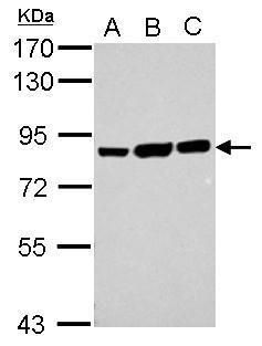
- Western blot analysis of Ezrin using 30 µg of A) 293T (B) A431 and C) HeLa lysate. Samples were loaded onto a 7.5% SDS-PAGE gel and probed with an Ezrin polyclonal antibody (Product # PA5-29358) at a dilution of 1:10,000.
- Submitted by
- Invitrogen Antibodies (provider)
- Main image

- Experimental details
- CRISPR-Cas9 mediated genome editing ofEzrin (as confirmed by next generation sequencing) was achieved by using LentiArray™ Lentiviral sgRNA (Product # A32042, AssayID CRISPR864181_LV) and LentiArray Cas9 Lentivirus (Product # A32064). Fig (a) Western blot analysis of Ezrin was performed by loading 30 µg of HeLa Wild Type (Lane 1), HeLa Cas9 (Lane 2) and HeLa Cas9 cells transduced with Ezrin Lentiviral sgRNA (Lane 3) whole cell extracts. The samples were electrophoresed using NuPAGE™ Novex™ 4-12% Bis-Tris Protein Gel (Product # NP0322BOX). Resolved proteins were then transferred onto a nitrocellulose membrane (Product # IB23001) by iBlot® 2 Dry Blotting System (Product # IB21001). The blot was probed with Anti-Ezrin Polyclonal Antibody (Product # PA5-29358) using 1:10,000 dilution and Goat anti-Rabbit IgG (H+L) Superclonal™ Recombinant Secondary Antibody, HRP (Product # A27036 1:8,000 dilution).Chemiluminescent detection was performed using Novex® ECL Chemiluminescent Substrate Reagent Kit (Product # WP20005). Loss of signal in sgRNA transduced cells using the LentiArray™ CRISPR product line confirms that antibody is specific toEzrin (Fig (b)).

- Submitted by
- Invitrogen Antibodies (provider)
- Main image

- Experimental details
- Western Blot using Ezrin Polyclonal Antibody (Product # PA5-29358). Various whole cell extracts (30 µg) were separated by 7.5% SDS-PAGE, and the membrane was blotted with Ezrin Polyclonal Antibody (Product # PA5-29358) diluted at 1:10,000. The HRP-conjugated anti-rabbit IgG antibody was used to detect the primary antibody.
- Submitted by
- Invitrogen Antibodies (provider)
- Main image

- Experimental details
- Knockdown of Ezrin was achieved by transfecting HCT 116 with Ezrin specific siRNAs (Silencer® select Product # s14796, s14795). Western blot analysis (Fig. a) was performed using Membrane enriched extracts from the Ezrin knockdown cells (lane 3), non-targeting scrambled siRNA transfected cells (lane 2) and untransfected cells (lane 1). The blot was probed with Ezrin Polyclonal Antibody (Product # PA5-29358, 1:10000 ) and Goat anti-Rabbit IgG (H+L) Superclonal™ Recombinant Secondary Antibody, HRP (Product # A27036, 1:4000). Densitometric analysis of this western blot is shown in histogram (Fig. b). Decrease in signal upon siRNA mediated knock down confirms that antibody is specific to Ezrin.

- Submitted by
- Invitrogen Antibodies (provider)
- Main image

- Experimental details
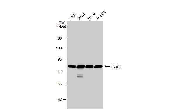
- Western blot was performed using Anti-Ezrin Polyclonal Antibody (Product # PA5-29358) and a ~90kDa band corresponding to Ezrin was observed across cell lines and tissues tested . Membrane enriched extracts (30 µg lysate) of HeLa (Lane 1), HCT 116 (Lane 2), A-431 (Lane 3), HaCaT (Lane 4), NTERA-2 cl.D1 (Lane 5), HEK-293 (Lane 6), Mouse Testis (Lane 7), Mouse Brain (Lane 8) were electrophoresed using NuPAGE™ 4-12% Bis-Tris Protein Gel (Product # NP0321BOX). Resolved proteins were then transferred onto a Nitrocellulose membrane (Product # LC2001) by iBlot® 2 Dry Blotting System (Product # IB21001). The blot was probed with the primary antibody (1:10000 dilution) and detected by chemiluminescence with Goat anti-Rabbit IgG (H+L) Superclonal™ Recombinant Secondary Antibody, HRP (Product # A27036, 1:4000) using the iBright FL 1000 (Product # A32752). Chemiluminescent detection was performed using Novex® ECL Chemiluminescent Substrate Reagent Kit (Product # WP20005).Another isoform around 70kDa was observed in some cell lines.

Supportive validation
- Submitted by
- Invitrogen Antibodies (provider)
- Main image

- Experimental details
- Immunocytochemistry-Immunofluorescence analysis of Ezrin was performed in HeLa cells fixed in 4% paraformaldehyde at RT for 15 min. Green: Ezrin Polyclonal Antibody (Product # PA5-29358) diluted at 1:1000. Red: alpha Tubulin, a cytoskeleton marker. Blue: Hoechst 33342 staining.

- Submitted by
- Invitrogen Antibodies (provider)
- Main image

- Experimental details
- Ezrin Polyclonal Antibody detects Ezrin protein at cytoplasm by immunofluorescent analysis. Sample: HCT116 cells were fixed in ice-cold MeOH for 5 min. Green: Ezrin protein stained by Ezrin Polyclonal Antibody (Product # PA5-29358) diluted at 1:1,000. Blue: Hoechst 33342 staining.

- Submitted by
- Invitrogen Antibodies (provider)
- Main image

- Experimental details
- Ezrin Polyclonal Antibody detects Ezrin protein at cell membrane by immunofluorescent analysis. Sample: HeLa cells were fixed in 4% paraformaldehyde at RT for 15 min. Green: Ezrin stained by Ezrin Polyclonal Antibody (Product # PA5-29358) diluted at 1:500. Red: alpha Tubulin, a cytoskeleton marker, stained by alpha Tubulin Polyclonal Antibody [GT114] (Product # MA5-31466) diluted at 1:1,000. Blue: Fluoroshield with DAPI .

Supportive validation
- Submitted by
- Invitrogen Antibodies (provider)
- Main image
- Experimental details
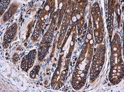
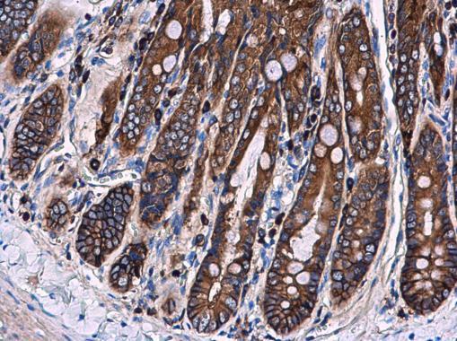
- Ezrin Polyclonal Antibody detects Ezrin protein at cytoplasm and membrane in rat duodenum by immunohistochemical analysis. Sample: Paraffin-embedded rat duodenum. Ezrin Polyclonal Antibody (Product # PA5-29358) diluted at 1:500. Antigen Retrieval: Citrate buffer, pH 6.0, 15 min.
- Submitted by
- Invitrogen Antibodies (provider)
- Main image
- Experimental details
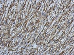
- Immunohistochemical analysis of paraffin-embedded C2C12 xenograft, using Ezrin (Product # PA5-29358) antibody at 1:500 dilution. Antigen Retrieval: EDTA based buffer, pH 8.0, 15 min.
- Submitted by
- Invitrogen Antibodies (provider)
- Main image

- Experimental details
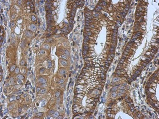
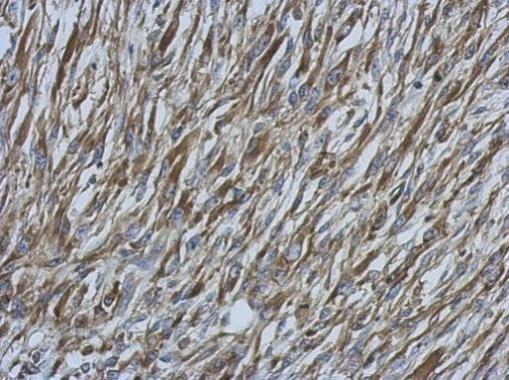
- Immunohistochemical analysis of paraffin-embedded human colon carcinoma, using Ezrin (Product # PA5-29358) antibody at 1:500 dilution. Antigen Retrieval: EDTA based buffer, pH 8.0, 15 min.