Explore
 Validate
 Learn
Learn Western blot
 Immunohistochemistry
 Blocking/Neutralizing
Antibody data
- Antibody Data
- Antigen structure
- References [1]
- Comments [0]
- Validations
- Immunohistochemistry [1]
- Flow cytometry [2]
- Other assay [1]
Submit
Validation data
Reference
Comment
Report error
- Product number
- PA5-47274 - Provider product page

- Provider
- Invitrogen Antibodies
- Product name
- Neuropilin 2 Polyclonal Antibody
- Antibody type
- Polyclonal
- Antigen
- Recombinant full-length protein
- Description
- In direct ELISAs, approximately 50% cross-reactivity with recombinant mouse Neuropilin-2 is observed, and approximately 25% cross-reactivity with recombinant rat (rr) Neuropilin-2 is observed and less than 5% cross-reactivity with rrNeuropilin-1 is observed. Reconstitute at 0.2 mg/mL in sterile PBS. Endoxin level is
- Reactivity
- Human, Mouse, Rat
- Host
- Goat
- Isotype
- IgG
- Vial size
- 100 μg
- Concentration
- 0.2 mg/mL
- Storage
- -20°C, Avoid Freeze/Thaw Cycles
Submitted references Smad3 Regulates Neuropilin 2 Transcription by Binding to its 5' Untranslated Region.
Xie X, Urabe G, Marcho L, Williams C, Guo LW, Kent KC
Journal of the American Heart Association 2020 Apr 21;9(8):e015487
Journal of the American Heart Association 2020 Apr 21;9(8):e015487
No comments: Submit comment
Supportive validation
- Submitted by
- Invitrogen Antibodies (provider)
- Main image

- Experimental details
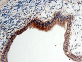
- Immunohistochemical analysis of Neuropilin 2 in immersion fixed paraffin-embedded sections of human pancreatic cancer tissue. Samples were incubated in Neuropilin 2 polyclonal antibody (Product # PA5-47274) using a dilution of 5 µg/mL overnight at 4 °C. Before incubation with the primary antibody, tissue was subjected to heat-induced epitope retrieval using Antigen Retrieval Reagent-basic. Tissue was stained using the Anti-Goat HRP-DAB Cell & Tissue Staining Kit (brown) and counterstained with hematoxylin (blue).
Supportive validation
- Submitted by
- Invitrogen Antibodies (provider)
- Main image

- Experimental details
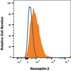
- Flow cytometry of Neuropilin 2 in HUVEC human umbilical vein endothelial cells. Samples were incubated in Neuropilin 2 polyclonal antibody (Product # PA5-47274) followed by Phycoerythrin-conjugated anti-Goat IgG.

- Submitted by
- Invitrogen Antibodies (provider)
- Main image
- Experimental details
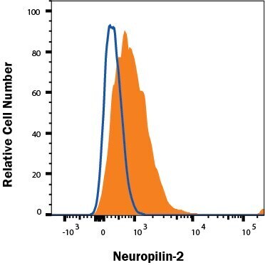
- Flow cytometry of Neuropilin 2 in HUVEC human umbilical vein endothelial cells. Samples were incubated in Neuropilin 2 polyclonal antibody (Product # PA5-47274) followed by Phycoerythrin-conjugated anti-Goat IgG.
Supportive validation
- Submitted by
- Invitrogen Antibodies (provider)
- Main image

- Experimental details
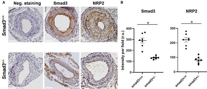
- Figure 5 Neuropilin 2 (NRP2) is reduced in arteries of Smad3-haploinsufficient (vs wild-type) mice. Wire injury experiments were performed with Smad3 +/+ and Smad3 +/- mice as described in the Methods section. Immunohistochemistry was performed on cross-sections of injured arteries collected 28 days after wire injury. A , Representative immunostained cross-sections. Negative staining: no primary antibody. Scale bar: 50 mum. B , Quantification of immunohistochemistry analysis. Colorimetric intensity (per image field) on a total of 7 cross-sections from 4 animals was averaged to calculate mean+-SEM (n=4 mice). Statistics: unpaired Student t test; * P