Explore
 Validate
 Learn
Learn Western blot
Antibody data
- Antibody Data
- Antigen structure
- References [0]
- Comments [0]
- Validations
- Western blot [2]
- Immunohistochemistry [1]
- Flow cytometry [1]
Submit
Validation data
Reference
Comment
Report error
- Product number
- ANR-062-200UL - Provider product page

- Provider
- Invitrogen Antibodies
- Product name
- Neuropilin-2 (NRP2) (extracellular) Polyclonal Antibody
- Antibody type
- Polyclonal
- Antigen
- Other
- Reactivity
- Human, Mouse, Rat
- Host
- Rabbit
- Isotype
- IgG
- Vial size
- 200 µL
- Concentration
- 0.8 mg/mL
- Storage
- -20° C, Avoid Freeze/Thaw Cycles
No comments: Submit comment
Supportive validation
- Submitted by
- Invitrogen Antibodies (provider)
- Main image
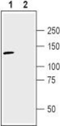
- Experimental details
- Western blot analysis of human U-87 MG glioma cell line lysate: - 1. Anti-Neuropilin-2 (NRP2) (extracellular) Antibody (#ANR-062), (1:200). 2. Anti-Neuropilin-2 (NRP2) (extracellular) Antibody , preincubated with Neuropilin-2/NRP2 (extracellular) Blocking Peptide (#BLP-NR062).
- Submitted by
- Invitrogen Antibodies (provider)
- Main image

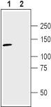
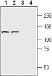
- Experimental details
- Western blot analysis of rat brain membranes (lanes 1 and 3) and mouse brain synaptosomal fraction (lanes 2 and 4): - 1,2. Anti-Neuropilin-2 (NRP2) (extracellular) Antibody (#ANR-062), (1:200).3,4. Anti-Neuropilin-2 (NRP2) (extracellular) Antibody , preincubated with Neuropilin-2/NRP2 (extracellular) Blocking Peptide (#BLP-NR062).
Supportive validation
- Submitted by
- Invitrogen Antibodies (provider)
- Main image
- Experimental details
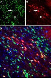
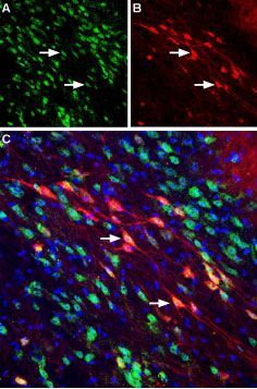
- Expression of Neuropilin-2 in mouse substantia nigra pars compacta - Immunohistochemical staining of perfusion-fixed frozen mouse brain sections using Anti-Neuropilin-2 (NRP2) (extracellular) Antibody (#ANR-062), (1:400). A. NRP2 staining (green) reveals several neuronal profiles of NRP2 positive neurons that are distributed along the substantia nigra pars compacta. B. The same section stained for Calbindin-D28K (red) shows positive neurons along the substantia nigra pars compacta. C. Merge of the two images reveals co-localization in several neurons (arrows). DAPI counterstain is used to visualize nuclei of all cells (blue).
Supportive validation
- Submitted by
- Invitrogen Antibodies (provider)
- Main image
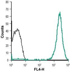
- Experimental details
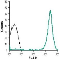
- Cell surface detection of Neuropilin-2 in live intact human THP-1 acute monocytic leukemia cell line: - (black) Unstained cells + goat Anti-rabbit-AlexaFluor-647 secondary Antibody . (green) Cells + Anti-Neuropilin-2 (NRP2) (extracellular) Antibody (#ANR-062), (1:15) + goat Anti-rabbit-AlexaFluor-647 secondary Antibody .