Explore
 Validate
 Learn
Learn Western blot
 Immunocytochemistry
Antibody data
- Antibody Data
- Antigen structure
- References [5]
- Comments [0]
- Validations
- Western blot [5]
- Immunocytochemistry [1]
- Immunoprecipitation [1]
- Immunohistochemistry [1]
Submit
Validation data
Reference
Comment
Report error
- Product number
- GTX100277 - Provider product page

- Provider
- GeneTex
- Proper citation
- GeneTex Cat#GTX100277, RRID:AB_1240835
- Product name
- FOXO3A antibody [C3], C-term
- Antibody type
- Polyclonal
- Reactivity
- Human, Mouse
- Host
- Rabbit
Submitted references Aβ exacerbates α-synuclein-induced neurotoxicity through impaired insulin signaling in α-synuclein-overexpressed human SK-N-MC neuronal cells.
Transcriptional activation of muscle atrophy promotes cardiac muscle remodeling during mammalian hibernation.
Sp1-mediated microRNA-182 expression regulates lung cancer progression.
Quercetin 3-O-methyl ether protects FL83B cells from copper induced oxidative stress through the PI3K/Akt and MAPK/Erk pathway.
The protective effects of spirulina in cyclophosphamide induced nephrotoxicity and urotoxicity in rats.
Chang CC, Li HH, Chang YT, Ho YJ, Hsieh LJ, Chiu PY, Cheng YS, Lin CL, Lai TJ
CNS neuroscience & therapeutics 2018 Jan;24(1):47-57
CNS neuroscience & therapeutics 2018 Jan;24(1):47-57
Transcriptional activation of muscle atrophy promotes cardiac muscle remodeling during mammalian hibernation.
Zhang Y, Aguilar OA, Storey KB
PeerJ 2016;4:e2317
PeerJ 2016;4:e2317
Sp1-mediated microRNA-182 expression regulates lung cancer progression.
Yang WB, Chen PH, Hsu T 1st, Fu TF, Su WC, Liaw H, Chang WC, Hung JJ
Oncotarget 2014 Feb 15;5(3):740-53
Oncotarget 2014 Feb 15;5(3):740-53
Quercetin 3-O-methyl ether protects FL83B cells from copper induced oxidative stress through the PI3K/Akt and MAPK/Erk pathway.
Tseng HL, Li CJ, Huang LH, Chen CY, Tsai CH, Lin CN, Hsu HY
Toxicology and applied pharmacology 2012 Oct 1;264(1):104-13
Toxicology and applied pharmacology 2012 Oct 1;264(1):104-13
The protective effects of spirulina in cyclophosphamide induced nephrotoxicity and urotoxicity in rats.
Sinanoglu O, Yener AN, Ekici S, Midi A, Aksungar FB
Urology 2012 Dec;80(6):1392.e1-6
Urology 2012 Dec;80(6):1392.e1-6
No comments: Submit comment
Enhanced validation
Supportive validation
- Submitted by
- GeneTex (provider)
- Enhanced method
- Genetic validation
- Main image
- Experimental details
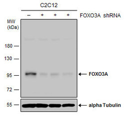
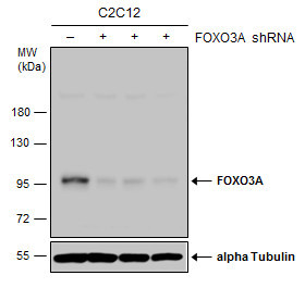
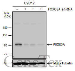
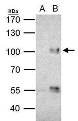
- Non-transfected (¡V) and transfected (+) C2C12 whole cell extracts (30 ?g) were separated by 7.5% SDS-PAGE, and the membrane was blotted with FOXO3A antibody [C3], C-term (GTX100277) diluted at 1:500. The HRP-conjugated anti-rabbit IgG antibody (GTX213110-01) was used to detect the primary antibody.
Supportive validation
- Submitted by
- GeneTex (provider)
- Main image

- Experimental details
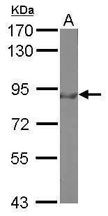
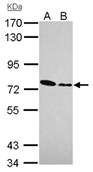
- Sample (30 ?g of whole cell lysate) A: 293T 7.5% SDS PAGE GTX100277 diluted at 1:500 The HRP-conjugated anti-rabbit IgG antibody (GTX213110-01) was used to detect the primary antibody.

- Submitted by
- GeneTex (provider)
- Main image

- Experimental details
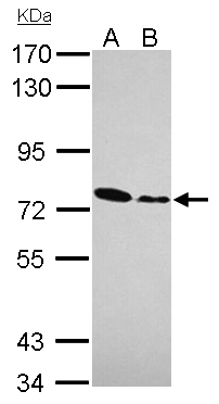
- Sample (50 ?g of whole cell lysate) A: Mouse brain 7.5% SDS PAGE GTX100277 diluted at 1:500 The HRP-conjugated anti-rabbit IgG antibody (GTX213110-01) was used to detect the primary antibody.
- Submitted by
- GeneTex (provider)
- Main image
- Experimental details
- Sample (30 ?g of whole cell lysate) A: Jurkat B: Raji 7.5% SDS PAGE GTX100277 diluted at 1:1000 The HRP-conjugated anti-rabbit IgG antibody (GTX213110-01) was used to detect the primary antibody.
- Submitted by
- GeneTex (provider)
- Main image
- Experimental details
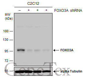
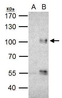
- Non-transfected (¡V) and transfected (+) C2C12 whole cell extracts (30 ?g) were separated by 7.5% SDS-PAGE, and the membrane was blotted with FOXO3A antibody [C3], C-term (GTX100277) diluted at 1:500. The HRP-conjugated anti-rabbit IgG antibody (GTX213110-01) was used to detect the primary antibody.
Supportive validation
- Submitted by
- GeneTex (provider)
- Main image
- Experimental details
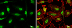
- FOXO3A antibody [C3], C-term detects FOXO3A protein at nucleus by immunofluorescent analysis.Sample: HeLa cells were fixed in 4% paraformaldehyde at RT for 15 min.Green: FOXO3A protein stained by FOXO3A antibody [C3], C-term (GTX100277) diluted at 1:500.Red: phalloidin, a cytoskeleton marker, diluted at 1:200.Scale bar = 10 £gm.

Supportive validation
- Submitted by
- GeneTex (provider)
- Main image
- Experimental details
- FOXO3A antibody immunoprecipitates FOXO3A protein in IP experiments. IP Sample: Jurkat whole cell lysate/extract A. Control with 2 £gg of preimmune rabbit IgG B. Immunoprecipitation of FOXO3A protein by 2 £gg of FOXO3A antibody (GTX101150) 5% SDS-PAGE The immunoprecipitated FOXO3A protein was detected by FOXO3A antibody (GTX101150) diluted at 1:1000. EasyBlot anti-rabbit IgG (GTX221666-01) was used as a secondary reagent.
Supportive validation
- Submitted by
- GeneTex (provider)
- Main image
- Experimental details
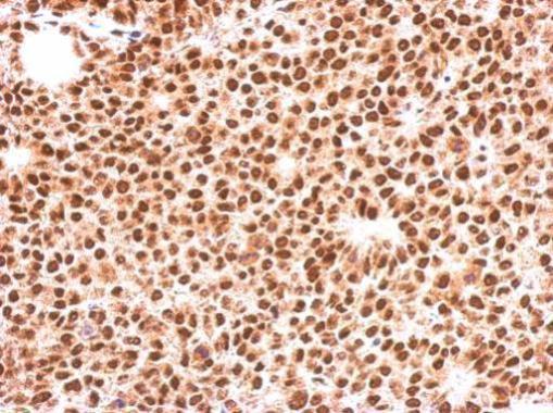
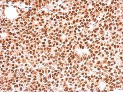
- Immunohistochemical analysis of paraffin-embedded Huh-7 xenograft, using FOXO3A(GTX100277) antibody at 1:500 dilution.