Explore
 Validate
 Learn
Learn Western blot
 Immunocytochemistry
Antibody data
- Antibody Data
- Antigen structure
- References [3]
- Comments [0]
- Validations
- Immunocytochemistry [2]
- Flow cytometry [1]
- Chromatin Immunoprecipitation [2]
- Other assay [1]
Submit
Validation data
Reference
Comment
Report error
- Product number
- 720128 - Provider product page

- Provider
- Invitrogen Antibodies
- Product name
- FOXO3A Polyclonal Antibody
- Antibody type
- Polyclonal
- Antigen
- Synthetic peptide
- Description
- These Polyclonal antibodies are of rabbit origin developed by immunizing animals with proteins or peptides. The polyclonal antibody is purified by affinity purification from the rabbit sera generated after immunizing the rabbits with a specific type of protein or peptide. The purified antibody is tested for its functionality in various relevant research applications. The antibody is developed for Research Use Only and is non-hazardous or non-infectious in nature. This antibody is predicted to react with Monkey, Goat, Pig, Bovine and Rabbit.
- Reactivity
- Human, Mouse
- Host
- Rabbit
- Isotype
- IgG
- Vial size
- 100 μg
- Concentration
- 0.5 mg/mL
- Storage
- Store at 4°C short term. For long term storage, store at -20°C, avoiding freeze/thaw cycles.
Submitted references FoxO3a Inhibits Tamoxifen-Resistant Breast Cancer Progression by Inducing Integrin α5 Expression.
The neuroprotective mechanism of lithium after ischaemic stroke.
Etv6 activates vegfa expression through positive and negative transcriptional regulatory networks in Xenopus embryos.
Ricci E, Fava M, Rizza P, Pellegrino M, Bonofiglio D, Casaburi I, Lanzino M, Giordano C, Bruno R, Sirianni R, Barone I, Sisci D, Morelli C
Cancers 2022 Jan 2;14(1)
Cancers 2022 Jan 2;14(1)
The neuroprotective mechanism of lithium after ischaemic stroke.
Chen B, Zhang M, Ji M, Zhang D, Chen B, Gong W, Li X, Zhou Y, Dong C, Wen G, Zhan X, Wu X, Yuan H, Xu E, Xia M, Verkhratsky A, Li B
Communications biology 2022 Feb 3;5(1):105
Communications biology 2022 Feb 3;5(1):105
Etv6 activates vegfa expression through positive and negative transcriptional regulatory networks in Xenopus embryos.
Li L, Rispoli R, Patient R, Ciau-Uitz A, Porcher C
Nature communications 2019 Mar 6;10(1):1083
Nature communications 2019 Mar 6;10(1):1083
No comments: Submit comment
Supportive validation
- Submitted by
- Invitrogen Antibodies (provider)
- Main image

- Experimental details
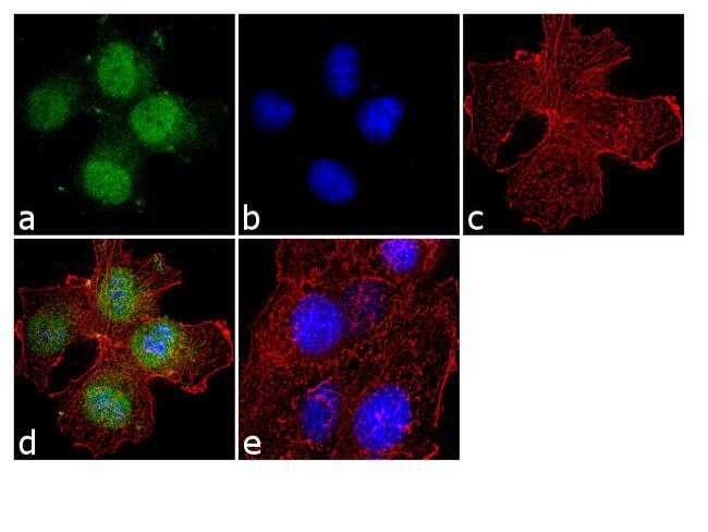
- Immunofluorescence was performed on fixed and permeabilized HeLa cells for detection of FOXO3A using Anti-FOXO3A Rabbit Polyclonal Antibody (Product # 720128, 2 µg/mL) and labeled with Goat anti-Rabbit IgG (H+L) Superclonal™ Secondary Antibody, Alexa Fluor® 488 conjugate (Product # A27034, 1:2000). Panel a) shows representative cells that were stained for detection and localization of FOXO3A protein (green), Panel b) is stained for nuclei (blue) using SlowFade® Gold Antifade Mountant with DAPI (Product # S36938). Panel c) represents cytoskeletal F-actin staining using Alexa Fluor® 555 Rhodamine Phalloidin (Product # R415, 1:300). Panel d) is a composite image of Panels a, b and c clearly demonstrating nuclear localization of FOXO3A. Panel e) represents control cells with no primary Antibody to assess background.

- Submitted by
- Invitrogen Antibodies (provider)
- Main image

- Experimental details
- Immunofluorescence was performed on fixed and permeabilized HeLa cells for detection of FOXO3A using Anti-FOXO3A Rabbit Polyclonal Antibody (Product # 720128, 2 µg/mL) and labeled with Goat anti-Rabbit IgG (Heavy Chain) Superclonal™ Secondary Antibody, Alexa Fluor® 488 conjugate (Product # A27034, 1:2000). Panel a) shows representative cells that were stained for detection and localization of FOXO3A protein (green), Panel b) is stained for nuclei (blue) using SlowFade® Gold Antifade Mountant with DAPI (Product # S36938). Panel c) represents cytoskeletal F-actin staining using Alexa Fluor® 555 Rhodamine Phalloidin (Product # R415, 1:300). Panel d) is a composite image of Panels a, b and c clearly demonstrating nuclear localization of FOXO3A. Panel e) represents control cells with no primary Antibody to assess background.
Supportive validation
- Submitted by
- Invitrogen Antibodies (provider)
- Main image
- Experimental details
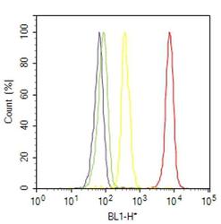
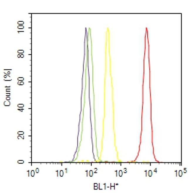
- Flow Cytometry analysis of FOXO3A was performed on Hela cells labeled with Anti-FOXO3A Rabbit Polyclonal Antibody (Product# 720128, 2- 4 ug/ 1M cells) or with rabbit isotype control and detected with Goat anti-Rabbit IgG (H+L) Superclonal™ Secondary Antibody, (Alexa Fluor® 488 conjugate, Product # A27034, 0.4 ug/ml, 1:2500) as represented by the red and yellow histograms respectively. The purple histogram represents unstained control cells and the green histogram represents no-primary-antibody control. A representative of 10,000 cells were acquired and analyzed for each sample using an Attune® Acoustic Focusing Cytometer (4468770).
Supportive validation
- Submitted by
- Invitrogen Antibodies (provider)
- Main image
- Experimental details
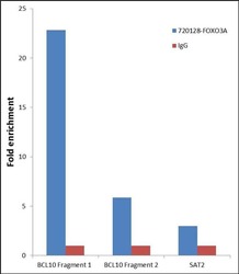
- Chromatin Immunoprecipitation (ChIP) was performed using Anti-FOXO3a Rabbit Polyclonal Antibody (Product # 720128, 3 µg) on sheared chromatin from 2 million Jurkat cells serum starved for 24 hours using the MAGnify ChIP system kit (Product # 49-2024). Normal Rabbit IgG was used as a negative IP control. The purified DNA was analyzed by 7500 Fast qPCR system (Product # 4351106) with optimized PCR primer pairs for the promoters of the active BCL10 region used as positive control target gene, and the region of the inactive SAT2 satellite repeat, used as negative control target gene. Data is presented as fold enrichment of the antibody signal versus the negative control IgG using the comparative CT method.

- Submitted by
- Invitrogen Antibodies (provider)
- Main image
- Experimental details
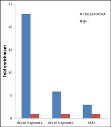
- Chromatin Immunoprecipitation (ChIP) was performed using Anti-FOXO3a Rabbit Polyclonal Antibody (Product # 720128, 3 µg) on sheared chromatin from 2 million Jurkat cells serum starved for 24 hours using the MAGnify ChIP system kit (Product # 49-2024). Normal Rabbit IgG was used as a negative IP control. The purified DNA was analyzed by 7500 Fast qPCR system (Product # 4351106) with optimized PCR primer pairs for the promoters of the active BCL10 region used as positive control target gene, and the region of the inactive SAT2 satellite repeat, used as negative control target gene. Data is presented as fold enrichment of the antibody signal versus the negative control IgG using the comparative CT method.

Supportive validation
- Submitted by
- Invitrogen Antibodies (provider)
- Main image

- Experimental details
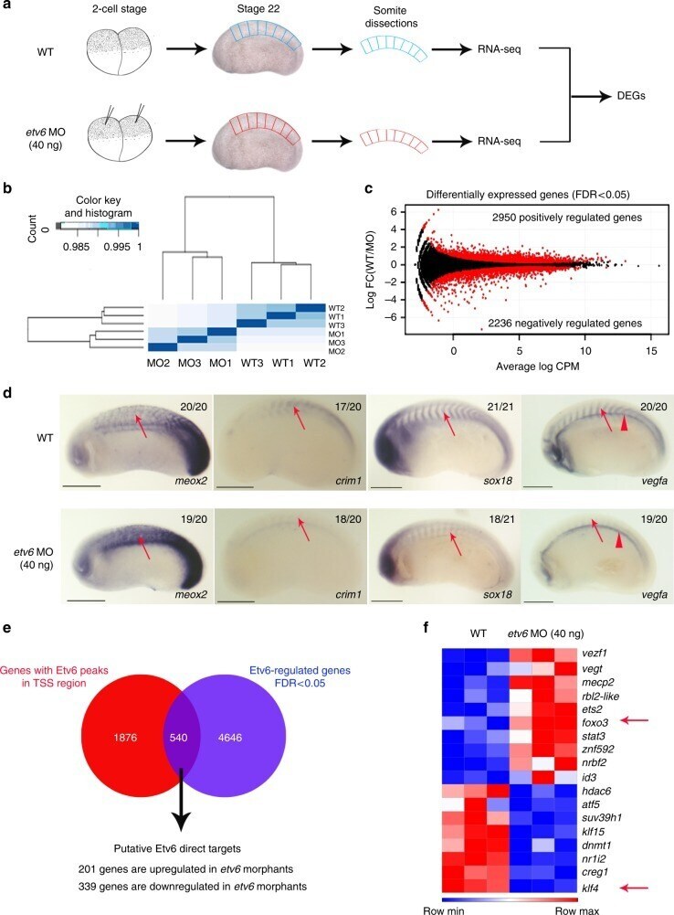
- Fig. 2 Identification of Etv6 direct transcriptional targets in the somites. a Experimental design. RNA-seq was performed on wild type (WT) and Etv6-deficient ( etv6 MO-injected embryos) somite explants dissected from stage 22 embryos. Differentially expressed genes (DEGs) were identified by comparing the transcriptome of these tissues. b Spearman correlation analysis on triple biological RNA-seq replicates. c MA plot (magnitude of the difference versus amplitude of the signal) showing the fold-change in gene expression of all genes in WT versus Etv6-deficient somites (log 2 FC) compared to their expression levels (log 2 CPM). Black dots, non-significant change; red dots, differentially expressed genes (DEGs). The number of positively and negatively regulated DEGs is indicated. d WISH showing the expression of DEGs in stage 22 WT and Etv6-deficient embryos. Meox2 expression is upregulated in the somites (arrows) in Etv6-deficient embryos whereas expression of crim1 , sox18 and vegfa is downregulated. Note that vegfa expression in the hypochord is unaffected (arrowheads). Embryos are shown in lateral view with anterior to the left and dorsal to the top. Numbers in top right corner indicate the number of embryos exhibiting the phenotype pictured (scale bars: 0.5 mm). e The intersection between DEGs and genes harbouring Etv6 ChIP-seq peaks in their TSS region reveals 540 putative direct target genes. f Foxo3 and klf4 (arrows), known transcriptional regul