Explore
 Validate
 Learn
Learn Western blot
 Immunocytochemistry
Antibody data
- Antibody Data
- Antigen structure
- References [2]
- Comments [0]
- Validations
- Immunocytochemistry [2]
- Immunohistochemistry [6]
- Other assay [1]
Submit
Validation data
Reference
Comment
Report error
- Product number
- PA5-27145 - Provider product page

- Provider
- Invitrogen Antibodies
- Product name
- FOXO3A Polyclonal Antibody
- Antibody type
- Polyclonal
- Antigen
- Synthetic peptide
- Description
- Recommended positive controls: 293T, Mouse brain, C2C12, Jurkat, Raji. Predicted reactivity: Mouse (100%), Rat (100%), Pig (100%), Sheep (100%), Bovine (100%). Store product as a concentrated solution. Centrifuge briefly prior to opening the vial.
- Reactivity
- Human, Mouse, Rat, Xenopus
- Host
- Rabbit
- Isotype
- IgG
- Vial size
- 100 μL
- Concentration
- 0.56 mg/mL
- Storage
- Store at 4°C short term. For long term storage, store at -20°C, avoiding freeze/thaw cycles.
Submitted references Lipid Droplet-Derived Monounsaturated Fatty Acids Traffic via PLIN5 to Allosterically Activate SIRT1.
Impact of first-line cancer treatment on the follicle quality in cryopreserved ovarian samples from girls and young women.
Najt CP, Khan SA, Heden TD, Witthuhn BA, Perez M, Heier JL, Mead LE, Franklin MP, Karanja KK, Graham MJ, Mashek MT, Bernlohr DA, Parker L, Chow LS, Mashek DG
Molecular cell 2020 Feb 20;77(4):810-824.e8
Molecular cell 2020 Feb 20;77(4):810-824.e8
Impact of first-line cancer treatment on the follicle quality in cryopreserved ovarian samples from girls and young women.
Pampanini V, Wagner M, Asadi-Azarbaijani B, Oskam IC, Sheikhi M, Sjödin MOD, Lindberg J, Hovatta O, Sahlin L, Björvang RD, Otala M, Damdimopoulou P, Jahnukainen K
Human reproduction (Oxford, England) 2019 Sep 29;34(9):1674-1685
Human reproduction (Oxford, England) 2019 Sep 29;34(9):1674-1685
No comments: Submit comment
Supportive validation
- Submitted by
- Invitrogen Antibodies (provider)
- Main image

- Experimental details
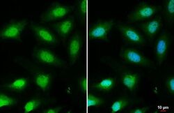
- Immunocytochemistry-Immunofluorescence analysis of FOXO3A was performed in HeLa cells fixed in 4% paraformaldehyde at RT for 15 min. Green: FOXO3A Polyclonal Antibody (Product # PA5-27145) diluted at 1:1000. Blue: Hoechst 33342 staining. Scale bar = 10 µm.

- Submitted by
- Invitrogen Antibodies (provider)
- Main image
- Experimental details
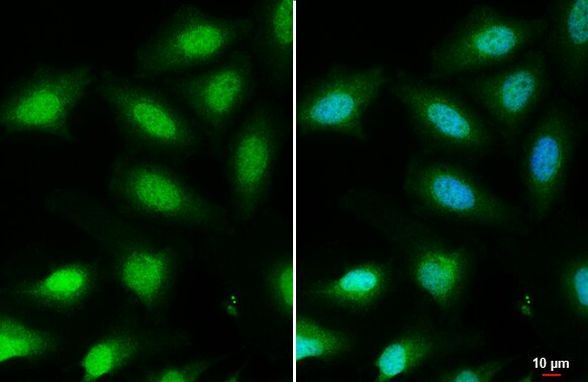
- Immunocytochemistry-Immunofluorescence analysis of FOXO3A was performed in HeLa cells fixed in 4% paraformaldehyde at RT for 15 min. Green: FOXO3A Polyclonal Antibody (Product # PA5-27145) diluted at 1:1000. Blue: Hoechst 33342 staining. Scale bar = 10 µm.
Supportive validation
- Submitted by
- Invitrogen Antibodies (provider)
- Main image
- Experimental details
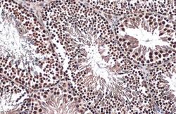
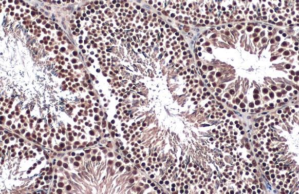
- FOXO3A Polyclonal Antibody detects FOXO3A protein at nucleus by immunohistochemical analysis. Sample: Paraffin-embedded mouse testis. FOXO3A stained by FOXO3A Polyclonal Antibody (Product # PA5-27145) diluted at 1:500. Antigen Retrieval: Citrate buffer, pH 6.0, 15 min.
- Submitted by
- Invitrogen Antibodies (provider)
- Main image
- Experimental details
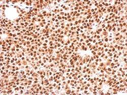
- Immunohistochemical analysis of paraffin-embedded Huh-7 xenograft, using FOXO3A (Product # PA5-27145) antibody at 1:500 dilution. Antigen Retrieval: EDTA based buffer, pH 8.0, 15 min.
- Submitted by
- Invitrogen Antibodies (provider)
- Main image
- Experimental details
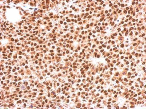
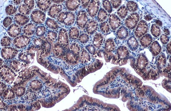
- FOXO3A Polyclonal Antibody detects FOXO3A protein at cytoplasm and nucleus by immunohistochemical analysis. Sample: Paraffin-embedded mouse duodenum. FOXO3A stained by FOXO3A Polyclonal Antibody (Product # PA5-27145) diluted at 1:2,000. Antigen Retrieval: Citrate buffer, pH 6.0, 15 min.
- Submitted by
- Invitrogen Antibodies (provider)
- Main image

- Experimental details
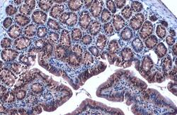
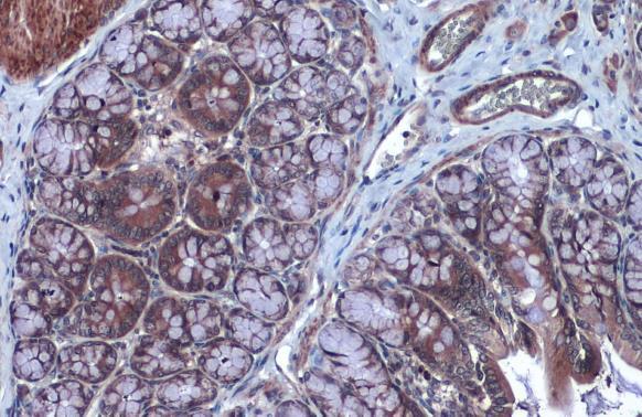
- FOXO3A Polyclonal Antibody detects FOXO3A protein at cytoplasm by immunohistochemical analysis. Sample: Paraffin-embedded rat colon. FOXO3A stained by FOXO3A Polyclonal Antibody (Product # PA5-27145) diluted at 1:2,000. Antigen Retrieval: Citrate buffer, pH 6.0, 15 min.
- Submitted by
- Invitrogen Antibodies (provider)
- Main image
- Experimental details
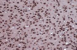
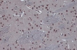
- FOXO3A Polyclonal Antibody detects FOXO3A protein at cytoplasm and nucleus by immunohistochemical analysis. Sample: Paraffin-embedded mouse brain. FOXO3A stained by FOXO3A Polyclonal Antibody (Product # PA5-27145) diluted at 1:2,000. Antigen Retrieval: Citrate buffer, pH 6.0, 15 min.
- Submitted by
- Invitrogen Antibodies (provider)
- Main image
- Experimental details
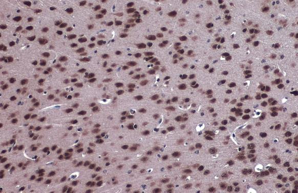
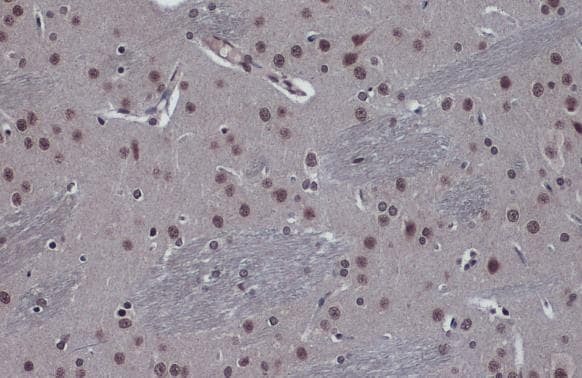
- FOXO3A Polyclonal Antibody detects FOXO3A protein at cytoplasm and nucleus by immunohistochemical analysis. Sample: Paraffin-embedded rat brain. FOXO3A stained by FOXO3A Polyclonal Antibody (Product # PA5-27145) diluted at 1:2,000. Antigen Retrieval: Citrate buffer, pH 6.0, 15 min.
Supportive validation
- Submitted by
- Invitrogen Antibodies (provider)
- Main image
- Experimental details
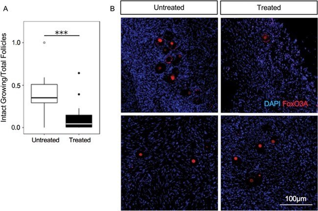
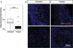
- Figure 4 Exposure to chemotherapy before ovarian tissue cryopreservation does not increase activation of ovarian follicles . ( A ) Box plots showing a significantly smaller proportion of growing follicles in ovarian tissue of treated ( n = 14) and untreated ( n = 13) patients. For each patient, the number of intact growing (intermediary and primary) follicles was divided by the total number of intact and atretic follicles (primordial, intermediary and primary) *** ( P < 0.001). ( B ) Immunofluorescence of ovarian tissue from a subset of treated ( n = 3) and untreated ( n = 5) patients labeled with fork head box O3A (FOXO3A) antibody shows no differences between the two groups. FOXO3A is located in the nuclei of oocytes and granulosa cells of both treated and untreated patients indicating non-activated follicles. Representative images of ovarian tissue sections are shown.