Explore
 Validate
 Learn
Learn Western blot
 Immunocytochemistry
Antibody data
- Antibody Data
- Antigen structure
- References [4]
- Comments [0]
- Validations
- Immunocytochemistry [4]
- Immunohistochemistry [3]
- Other assay [4]
Submit
Validation data
Reference
Comment
Report error
- Product number
- PA5-28228 - Provider product page

- Provider
- Invitrogen Antibodies
- Product name
- FDPS Polyclonal Antibody
- Antibody type
- Polyclonal
- Antigen
- Recombinant full-length protein
- Description
- Recommended positive controls: A549, H1299, HCT116, Mouse brain, rat liver. Predicted reactivity: Mouse (89%), Rat (89%), Pig (89%), Bovine (91%). Store product as a concentrated solution. Centrifuge briefly prior to opening the vial.
- Reactivity
- Human, Mouse, Rat
- Host
- Rabbit
- Isotype
- IgG
- Vial size
- 100 μL
- Concentration
- 1.35 mg/mL
- Storage
- Store at 4°C short term. For long term storage, store at -20°C, avoiding freeze/thaw cycles.
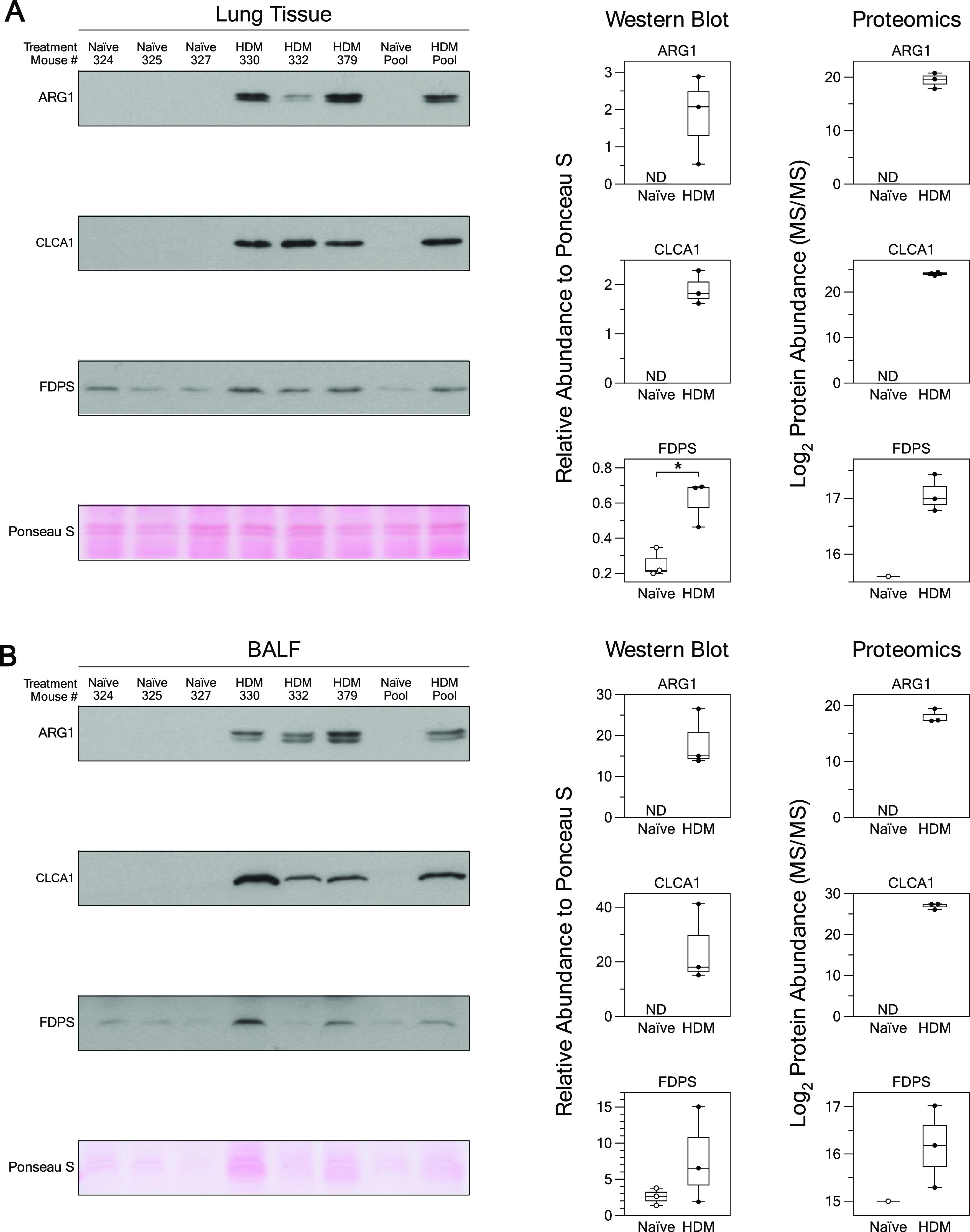
Submitted references Integrating Proteomes for Lung Tissues and Lavage Reveals Pathways That Link Responses in Allergen-Challenged Mice.
Regeneration of Cochlear Synapses by Systemic Administration of a Bisphosphonate.
FDPS promotes glioma growth and macrophage recruitment by regulating CCL20 via Wnt/β-catenin signalling pathway.
Butyrophilin 3A/CD277-Dependent Activation of Human γδ T Cells: Accessory Cell Capacity of Distinct Leukocyte Populations.
Mahood TH, Pascoe CD, Karakach TK, Jha A, Basu S, Ezzati P, Spicer V, Mookherjee N, Halayko AJ
ACS omega 2021 Jan 19;6(2):1171-1189
ACS omega 2021 Jan 19;6(2):1171-1189
Regeneration of Cochlear Synapses by Systemic Administration of a Bisphosphonate.
Seist R, Tong M, Landegger LD, Vasilijic S, Hyakusoku H, Katsumi S, McKenna CE, Edge ASB, Stankovic KM
Frontiers in molecular neuroscience 2020;13:87
Frontiers in molecular neuroscience 2020;13:87
FDPS promotes glioma growth and macrophage recruitment by regulating CCL20 via Wnt/β-catenin signalling pathway.
Chen Z, Chen G, Zhao H
Journal of cellular and molecular medicine 2020 Aug;24(16):9055-9066
Journal of cellular and molecular medicine 2020 Aug;24(16):9055-9066
Butyrophilin 3A/CD277-Dependent Activation of Human γδ T Cells: Accessory Cell Capacity of Distinct Leukocyte Populations.
Nerdal PT, Peters C, Oberg HH, Zlatev H, Lettau M, Quabius ES, Sousa S, Gonnermann D, Auriola S, Olive D, Määttä J, Janssen O, Kabelitz D
Journal of immunology (Baltimore, Md. : 1950) 2016 Oct 15;197(8):3059-3068
Journal of immunology (Baltimore, Md. : 1950) 2016 Oct 15;197(8):3059-3068
No comments: Submit comment
Supportive validation
- Submitted by
- Invitrogen Antibodies (provider)
- Main image

- Experimental details
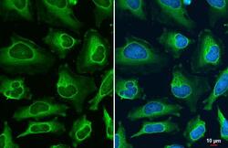
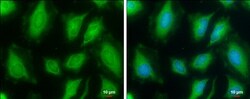
- Immunocytochemistry-Immunofluorescence analysis of FDPS was performed in HeLa cells fixed in ice-cold MeOH for 5 min. Green: FDPS Polyclonal Antibody (Product # PA5-28228) diluted at 1:500. Blue: Hoechst 33342 staining. Scale bar = 10 µm.

- Submitted by
- Invitrogen Antibodies (provider)
- Main image
- Experimental details
- FDPS Polyclonal Antibody detects FDPS protein at cytoplasm by immunofluorescent analysis. Sample: HeLa cells were fixed in ice-cold MeOH for 5 min. Green: FDPS stained by FDPS Polyclonal Antibody (Product # PA5-28228) diluted at 1:500.

- Submitted by
- Invitrogen Antibodies (provider)
- Main image

- Experimental details
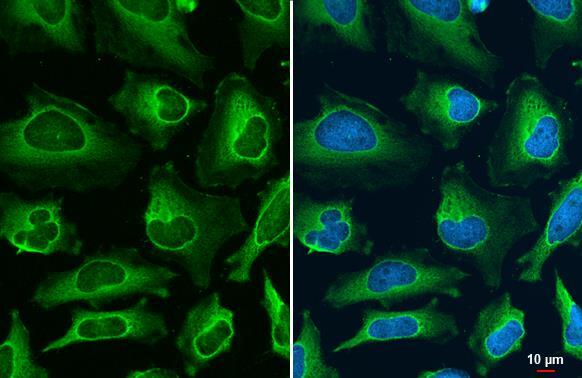
- FDPS Polyclonal Antibody detects FDPS protein at cytoplasm by immunofluorescent analysis. Sample: HeLa cells were fixed in ice-cold MeOH for 5 min. Green: FDPS stained by FDPS Polyclonal Antibody (Product # PA5-28228) diluted at 1:500.
- Submitted by
- Invitrogen Antibodies (provider)
- Main image
- Experimental details
- Immunocytochemistry-Immunofluorescence analysis of FDPS was performed in HeLa cells fixed in ice-cold MeOH for 5 min. Green: FDPS Polyclonal Antibody (Product # PA5-28228) diluted at 1:500. Blue: Hoechst 33342 staining. Scale bar = 10 µm.

Supportive validation
- Submitted by
- Invitrogen Antibodies (provider)
- Main image
- Experimental details
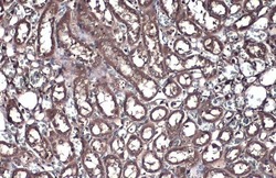
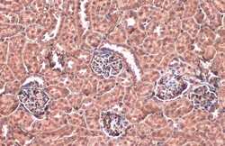
- FDPS Polyclonal Antibody detects FDPS protein at cytoplasm by immunohistochemical analysis. Sample: Paraffin-embedded rat kidney. FDPS stained by FDPS Polyclonal Antibody (Product # PA5-28228) diluted at 1:500. Antigen Retrieval: Citrate buffer, pH 6.0, 15 min.
- Submitted by
- Invitrogen Antibodies (provider)
- Main image
- Experimental details
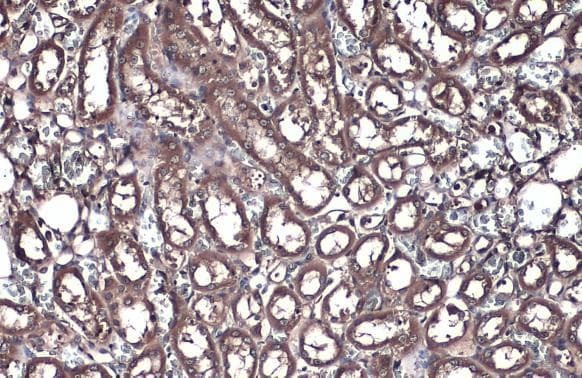
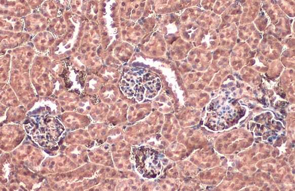
- FDPS Polyclonal Antibody detects FDPS protein at cytoplasm by immunohistochemical analysis. Sample: Paraffin-embedded mouse kidney. FDPS stained by FDPS Polyclonal Antibody (Product # PA5-28228) diluted at 1:500. Antigen Retrieval: Citrate buffer, pH 6.0, 15 min.
- Submitted by
- Invitrogen Antibodies (provider)
- Main image
- Experimental details
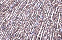
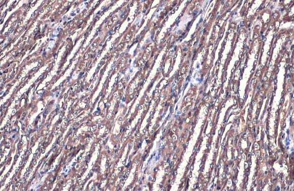
- Immunohistochemistry (Paraffin) analysis of FDPS was performed in paraffin-embedded mouse kidney tissue using FDPS Polyclonal Antibody (Product # PA5-28228) at a dilution of 1:500. Antigen Retrieval: Citrate buffer, pH 6.0, 15 min.
Supportive validation
- Submitted by
- Invitrogen Antibodies (provider)
- Main image

- Experimental details
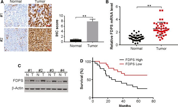
- FIGURE 1 FDPS is expressed at high levels in glioma tissues. The expression of FDPS in glioma tissue and adjacent non-tumour tissue from same patient was assessed by immunohistochemistry (A), real-time PCR (B) and Western blotting (C). (D) Kaplan-Meier curves showing the overall survival of glioma patients with low versus high FDPS expression ( P = 0.025). Error bars represent the SD, ** P < 0.01 (Scale bars, 50 mum)
- Submitted by
- Invitrogen Antibodies (provider)
- Main image
- Experimental details
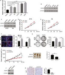
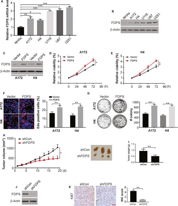
- FIGURE 2 FDPS regulates glioma cell growth. A, The protein expression levels of FDPS in human NHAs and glioma cell lines determined by real-time PCR. B, The protein expression levels of FDPS in human NHAs and glioma cell lines determined by Western blotting. C, A172 and H4 cells with FDPS overexpression were established. The level of FDPS in these established cell lines was verified by Western blotting. D and E, Cell proliferation was examined by MTS in A172 and H4 cells with FDPS overexpression. F, EdU assay of indicated cells with FDPS overexpression. G, Colony formation assays of indicated cells with FDPS overexpression. H, U87 cells with stable expression of shCon/shFDPS were subcutaneous injected into NSG mice. Tumour size was measured every 2 days. I, The weight of tumours formed at day 17th. J, The FDPS expression in tumours was analysed by Western blotting. K, The expression of Ki67 was evaluated by IHC staining. Error bars represent the SD, * P < 0.05; ** P < 0.01; *** P < 0.001 (Scale bars, 50 mum)
- Submitted by
- Invitrogen Antibodies (provider)
- Main image
- Experimental details
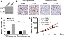
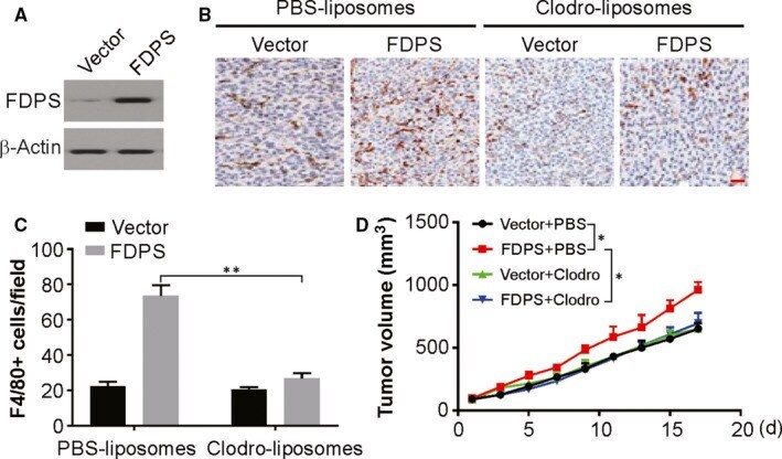
- FIGURE 4 FDPS promotes tumour growth in a macrophage-dependent manner. A, The level of FDPS in GL261 cells was analysed by Western blotting. B and C, Representative immunohistochemistry of F4/80+ cells in sections from glioma tumours obtained from C57BL/6J mice treated with clodronate liposomes or PBS liposomes. D, Volume of GL261 tumours treated as indicated. Error bars represent the SD, * P < 0.05; ** P < 0.01 (Scale bars, 50 mum)
- Submitted by
- Invitrogen Antibodies (provider)
- Main image
- Experimental details
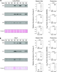
- Figure 2 Western blot validation of lung tissue and BALF proteomics. (A, B) Images of the scanned chemiluminescent film for ARG1, CLC1, and FDPS. Protein bands were normalized to total protein loading (Ponceau S) for quantification. Abbreviations used: BALF: bronchial alveolar lavage fluid, * (Welch's unpaired t -test p.adj