 Explore
 Validate
 Learn
Learn Western blot
Antibody data
- Antibody Data
- Antigen structure
- References [1]
- Comments [0]
- Validations
- Western blot [2]
- Immunocytochemistry [1]
- Immunohistochemistry [2]
Submit
Validation data
Reference
Comment
Report error
- Product number
- MAB7049 - Provider product page

- Provider
- R&D Systems
- Product name
- Human Isocitrate Dehydrogenase 1/IDH1 Antibody
- Antibody type
- Monoclonal
- Description
- Protein A or G purified from hybridoma culture supernatant. Detects human Isocitrate Dehydrogenase 1/IDH1 in ELISAs and Western blots.
- Reactivity
- Human
- Host
- Mouse
- Conjugate
- Unconjugated
- Antigen sequence
O75874- Isotype
- IgG
- Antibody clone number
- 843219
- Vial size
- 100 ug
- Concentration
- LYOPH
- Storage
- Use a manual defrost freezer and avoid repeated freeze-thaw cycles. 12 months from date of receipt, -20 to -70 °C as supplied. 1 month, 2 to 8 °C under sterile conditions after reconstitution. 6 months, -20 to -70 °C under sterile conditions after reconstitution.
Submitted references Mutant and Wild-Type Isocitrate Dehydrogenase 1 Share Enhancing Mechanisms Involving Distinct Tyrosine Kinase Cascades in Cancer.
Chen D, Xia S, Wang M, Lin R, Li Y, Mao H, Aguiar M, Famulare CA, Shih AH, Brennan CW, Gao X, Pan Y, Liu S, Fan J, Jin L, Song L, Zhou A, Mukherjee J, Pieper RO, Mishra A, Peng J, Arellano M, Blum WG, Lonial S, Boggon TJ, Levine RL, Chen J
Cancer discovery 2019 Jun;9(6):756-777
Cancer discovery 2019 Jun;9(6):756-777
No comments: Submit comment
Supportive validation
- Submitted by
- R&D Systems (provider)
- Main image

- Experimental details
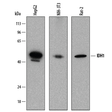
- Detection of Human, Mouse, and Rat Isocitrate Dehydrogenase 1/IDH1 by Western Blot. Western blot shows lysates of HepG2 human hepatocellular carcinoma cell line, NIH-3T3 mouse embryonic fibroblast cell line, and Rat-2 rat embryonic fibroblast cell line. PVDF membrane was probed with 0.25 µg/mL of Mouse Anti-Human Isocitrate Dehydrogenase 1/IDH1 Monoclonal Antibody (Catalog # MAB7049) followed by HRP-conjugated Anti-Mouse IgG Secondary Antibody (Catalog # HAF018). A specific band was detected for Isocitrate Dehydrogenase 1/IDH1 at approximately 46 kDa (as indicated). This experiment was conducted under reducing conditions and using Immunoblot Buffer Group 1.
- Submitted by
- R&D Systems (provider)
- Main image
- Experimental details
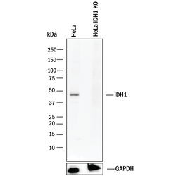
- Western Blot Shows Human Isocitrate Dehydrogenase 1/IDH1 Specificity by Using Knockout Cell Line. Western blot shows lysates of HeLa human cervical epithelial carcinoma parental cell line and Isocitrate Dehydrogenase 1/IDH1 knockout HeLa cell line (KO). PVDF membrane was probed with 0.25 µg/mL of Mouse Anti-Human Isocitrate Dehydrogenase 1/IDH1 Monoclonal Antibody (Catalog # MAB7049) followed by HRP-conjugated Anti-Mouse IgG Secondary Antibody (Catalog # HAF018). A specific band was detected for Isocitrate Dehydrogenase 1/IDH1 at approximately 46 kDa (as indicated) in the parental HeLa cell line, but is not detectable in knockout HeLa cell line. GAPDH (Catalog # MAB5718) is shown as a loading control. This experiment was conducted under reducing conditions and using Immunoblot Buffer Group 1.
Supportive validation
- Submitted by
- R&D Systems (provider)
- Main image

- Experimental details
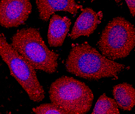
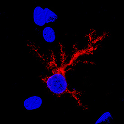
- Isocitrate Dehydrogenase 1/IDH1 in SK-BR-3 Human Cell Line. Isocitrate Dehydrogenase 1/IDH1 was detected in immersion fixed SK-BR-3 human breast cancer cell line using Mouse Anti-Human Isocitrate Dehydrogenase 1/IDH1 Monoclonal Antibody (Catalog # MAB7049) at 10 µg/mL for 3 hours at room temperature. Cells were stained using the NorthernLights™ 557-conjugated Anti-Mouse IgG Secondary Antibody (red; Catalog # NL007) and counterstained with DAPI (blue). Specific staining was localized to cytoplasm. View our protocol for Fluorescent ICC Staining of Cells on Coverslips.
Supportive validation
- Submitted by
- R&D Systems (provider)
- Main image
- Experimental details
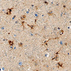
- Isocitrate Dehydrogenase 1/IDH1 in Human Brain. Isocitrate Dehydrogenase 1/IDH1 was detected in immersion fixed paraffin-embedded sections of human brain (cortex) using Mouse Anti-Human Isocitrate Dehydrogenase 1/IDH1 Monoclonal Antibody (Catalog # MAB7049) at 15 µg/mL overnight at 4 °C. Before incubation with the primary antibody, tissue was subjected to heat-induced epitope retrieval using Antigen Retrieval Reagent-Basic (Catalog # CTS013). Tissue was stained using the Anti-Mouse HRP-DAB Cell & Tissue Staining Kit (brown; Catalog # CTS002) and counterstained with hematoxylin (blue). Specific staining was localized to astrocytes. View our protocol for Chromogenic IHC Staining of Paraffin-embedded Tissue Sections.
- Submitted by
- R&D Systems (provider)
- Main image
- Experimental details
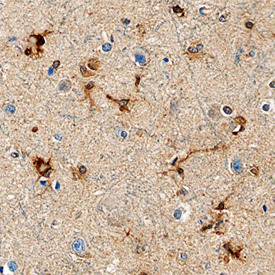
- Isocitrate Dehydrogenase 1/IDH1 in Rat Brain. Isocitrate Dehydrogenase 1/IDH1 was detected in perfusion fixed frozen sections of rat brain using Mouse Anti-Human Isocitrate Dehydrogenase 1/IDH1 Monoclonal Antibody (Catalog # MAB7049) at 15 µg/mL overnight at 4 °C. Tissue was stained using the NorthernLights™ 557-conjugated Anti-Mouse IgG Secondary Antibody (red; Catalog # NL007) and counterstained with DAPI (blue). Specific staining was localized to glial cell cytoplasm. View our protocol for Fluorescent IHC Staining of Frozen Tissue Sections.
