 Explore
 Validate
 Learn
Learn Western blot
 Immunoprecipitation
Antibody data
- Antibody Data
- Antigen structure
- References [1]
- Comments [0]
- Validations
- Immunoprecipitation [1]
- Immunohistochemistry [8]
- Other assay [1]
Submit
Validation data
Reference
Comment
Report error
- Product number
- PA5-28206 - Provider product page

- Provider
- Invitrogen Antibodies
- Product name
- IDH1 Polyclonal Antibody
- Antibody type
- Polyclonal
- Antigen
- Recombinant full-length protein
- Description
- Recommended positive controls: A431, HepG2, HeLa, Huh-7, Neuro2A , C8D30 , NIH-3T3 , Raw 264.7 , C2Cl2, DDDDk-tagged IDH1-transfected 293T. Predicted reactivity: Mouse (96%), Rat (97%), Zebrafish (85%), Xenopus laevis (88%), Sheep (95%), Rhesus Monkey (100%), Bovine (95%). Store product as a concentrated solution. Centrifuge briefly prior to opening the vial.
- Reactivity
- Human, Mouse, Rat
- Host
- Rabbit
- Isotype
- IgG
- Vial size
- 100 μL
- Concentration
- 0.69 mg/mL
- Storage
- Store at 4°C short term. For long term storage, store at -20°C, avoiding freeze/thaw cycles.
Submitted references Single-Cell Transcriptomic Atlas of Primate Ovarian Aging.
Wang S, Zheng Y, Li J, Yu Y, Zhang W, Song M, Liu Z, Min Z, Hu H, Jing Y, He X, Sun L, Ma L, Esteban CR, Chan P, Qiao J, Zhou Q, Izpisua Belmonte JC, Qu J, Tang F, Liu GH
Cell 2020 Feb 6;180(3):585-600.e19
Cell 2020 Feb 6;180(3):585-600.e19
No comments: Submit comment
Supportive validation
- Submitted by
- Invitrogen Antibodies (provider)
- Main image

- Experimental details
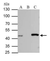
- IDH-1 antibody immunoprecipitates IDH-1 protein in IP experiments. IP Sample: HepG2 whole cell lysate/extract A : 30 µg whole cell lysate/extract of IDH1 protein expressing HepG2 cells B : Control with 2.5 µg of pre-immune rabbit IgG C : Immunoprecipitation of IDH-1 protein by 2.5 µg of IDH-1 antibody (Product # PA5-28206) 10% SDS-PAGE The immunoprecipitated IDH-1 protein was detected by IDH-1 antibody (Product # PA5-28206) diluted at 1:1,000. Anti-rabbit IgG (HRP) was used as a secondary reagent.

Supportive validation
- Submitted by
- Invitrogen Antibodies (provider)
- Main image
- Experimental details
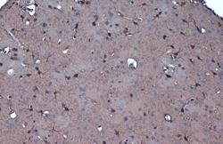
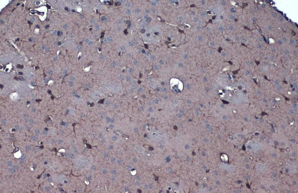
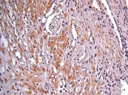
- IDH1 Polyclonal Antibody detects IDH1 protein at cytoplasm by immunohistochemical analysis. Sample: Paraffin-embedded mouse brain. IDH1 stained by IDH1 Polyclonal Antibody (Product # PA5-28206) diluted at 1:1,000. Antigen Retrieval: Citrate buffer, pH 6.0, 15 min.
- Submitted by
- Invitrogen Antibodies (provider)
- Main image
- Experimental details
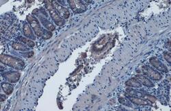
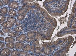
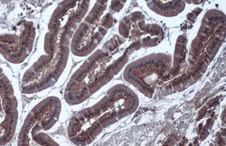
- IDH1 Polyclonal Antibody detects IDH1 protein at cytoplasm by immunohistochemical analysis. Sample: Paraffin-embedded mouse intestine. IDH1 stained by IDH1 Polyclonal Antibody (Product # PA5-28206) diluted at 1:1,000. Antigen Retrieval: Citrate buffer, pH 6.0, 15 min.
- Submitted by
- Invitrogen Antibodies (provider)
- Main image
- Experimental details
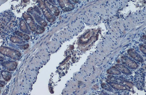
- IDH1 Polyclonal Antibody detects IDH1 protein at cytoplasm by immunohistochemical analysis. Sample: Paraffin-embedded rat colon. IDH1 stained by IDH1 Polyclonal Antibody (Product # PA5-28206) diluted at 1:1,000. Antigen Retrieval: Citrate buffer, pH 6.0, 15 min.
- Submitted by
- Invitrogen Antibodies (provider)
- Main image
- Experimental details
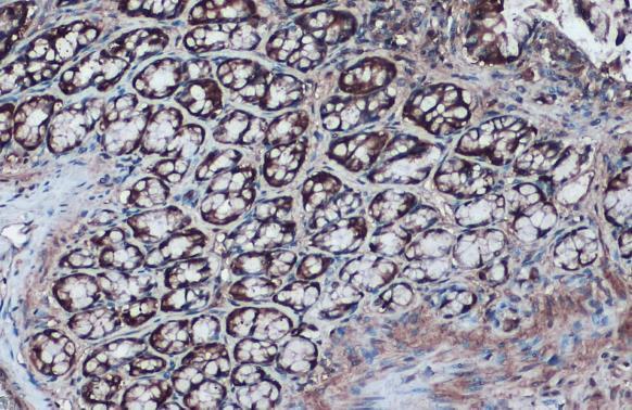
- Immunohistochemistry (Paraffin) analysis of IDH1 was performed in paraffin-embedded mouse duodenum tissue using IDH1 Polyclonal Antibody (Product # PA5-28206) at a dilution of 1:500.

- Submitted by
- Invitrogen Antibodies (provider)
- Main image
- Experimental details
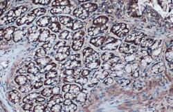
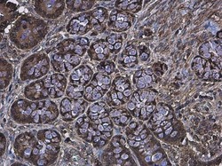
- Immunohistochemical analysis of paraffin-embedded human colon carcinoma, using IDH1 (Product # PA5-28206) antibody at 1:100 dilution. Antigen Retrieval: EDTA based buffer, pH 8.0, 15 min.

- Submitted by
- Invitrogen Antibodies (provider)
- Main image

- Experimental details
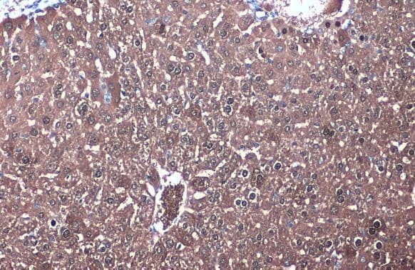
- IDH1 Polyclonal Antibody detects IDH1 protein at cytoplasm by immunohistochemical analysis. Sample: Paraffin-embedded mouse liver. IDH1 stained by IDH1 Polyclonal Antibody (Product # PA5-28206) diluted at 1:1,000. Antigen Retrieval: Citrate buffer, pH 6.0, 15 min.
- Submitted by
- Invitrogen Antibodies (provider)
- Main image
- Experimental details
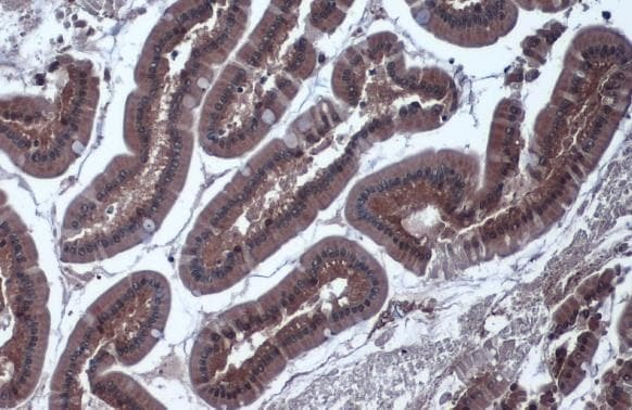
- IDH1 Polyclonal Antibody detects IDH1 protein at cytoplasm by immunohistochemical analysis. Sample: Paraffin-embedded rat duodenum. IDH1 stained by IDH1 Polyclonal Antibody (Product # PA5-28206) diluted at 1:1,000. Antigen Retrieval: Citrate buffer, pH 6.0, 15 min.
- Submitted by
- Invitrogen Antibodies (provider)
- Main image
- Experimental details
- Immunohistochemistry (Paraffin) analysis of IDH1 was performed in paraffin-embedded rat colon tissue using IDH1 Polyclonal Antibody (Product # PA5-28206) at a dilution of 1:500.

Supportive validation
- Submitted by
- Invitrogen Antibodies (provider)
- Main image
- Experimental details
- IDH-1 antibody immunoprecipitates IDH-1 protein in IP experiments. IP Sample: HepG2 whole cell lysate/extract A : 30 µg whole cell lysate/extract of IDH1 protein expressing HepG2 cells B : Control with 2.5 µg of pre-immune rabbit IgG C : Immunoprecipitation of IDH-1 protein by 2.5 µg of IDH-1 antibody (Product # PA5-28206) 10% SDS-PAGE The immunoprecipitated IDH-1 protein was detected by IDH-1 antibody (Product # PA5-28206) diluted at 1:1,000. Anti-rabbit IgG (HRP) was used as a secondary reagent.
