 Explore
 Validate
 Learn
Learn Western blot
 Immunocytochemistry
 Immunohistochemistry
Antibody data
- Antibody Data
- Antigen structure
- References [1]
- Comments [0]
- Validations
- Western blot [1]
- Immunocytochemistry [1]
Submit
Validation data
Reference
Comment
Report error
- Product number
- AMAb90578 - Provider product page

- Provider
- Atlas Antibodies
- Proper citation
- Atlas Antibodies Cat#AMAb90578, RRID:AB_2665593
- Product name
- Anti-IDH1
- Antibody type
- Monoclonal
- Description
- Monoclonal Antibody against Human IDH1, Clone ID: CL0219, Gene description: isocitrate dehydrogenase 1 (NADP+), soluble, Validated applications: ICC, IHC, WB, Uniprot ID: O75874, Storage: Store at +4°C for short term storage. Long time storage is recommended at -20°C.
- Reactivity
- Human
- Host
- Mouse
- Conjugate
- Unconjugated
- Isotype
- IgG
- Antibody clone number
- CL0219
- Vial size
- 100 µl
- Concentration
- 0.1 mg/ml
- Storage
- Store at +4°C for short term storage. Long time storage is recommended at -20°C.
- Handling
- The antibody solution should be gently mixed before use.
Submitted references Unravelling the proteomic landscape of extracellular vesicles in prostate cancer by density‐based fractionation of urine
Dhondt B, Geeurickx E, Tulkens J, Van Deun J, Vergauwen G, Lippens L, Miinalainen I, Rappu P, Heino J, Ost P, Lumen N, De Wever O, Hendrix A
Journal of Extracellular Vesicles 2020;9(1)
Journal of Extracellular Vesicles 2020;9(1)
No comments: Submit comment
Enhanced validation
- Submitted by
- Atlas Antibodies (provider)
- Enhanced method
- Genetic validation
- Main image
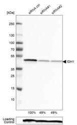
- Experimental details
- Western blot analysis in RT-4 cells transfected with control siRNA, target specific siRNA probe #1 and #2, using Anti-IDH1 antibody. Remaining relative intensity is presented. Loading control: Anti-PPIB.
- Sample type
- Human
- Protocol
- Protocol

Supportive validation
- Submitted by
- Atlas Antibodies (provider)
- Main image

- Experimental details
- Immunofluorescence staining of A-431 cells using the anti-IDH1 monoclonal antibody, showing specific staining in the cytosol and nuclear bodies in green. Microtubule- and nuclear probes are visualized in red and blue, respectively (where available).
- Sample type
- Human
