 Explore
 Validate
 Learn
LearnHPA001733
antibody from Atlas Antibodies
Targeting: DLG3
KIAA1232, MRX90, NE-Dlg, NEDLG, PPP1R82, SAP-102, SAP102
 Western blot
 Immunohistochemistry
Antibody data
- Antibody Data
- Antigen structure
- References [1]
- Comments [0]
- Validations
- Western blot [1]
Submit
Validation data
Reference
Comment
Report error
- Product number
- HPA001733 - Provider product page

- Provider
- Atlas Antibodies
- Proper citation
- Atlas Antibodies Cat#HPA001733, RRID:AB_1078675
- Product name
- Anti-DLG3
- Antibody type
- Polyclonal
- Description
- Polyclonal Antibody against Human DLG3, Gene description: discs, large homolog 3 (Drosophila), Alternative Gene Names: KIAA1232, MRX90, NE-Dlg, NEDLG, PPP1R82, SAP-102, SAP102, Validated applications: IHC, WB, Uniprot ID: Q92796, Storage: Store at +4°C for short term storage. Long time storage is recommended at -20°C.
- Reactivity
- Human
- Host
- Rabbit
- Conjugate
- Unconjugated
- Isotype
- IgG
- Vial size
- 100 µl
- Concentration
- 0.1 mg/ml
- Storage
- Store at +4°C for short term storage. Long time storage is recommended at -20°C.
- Handling
- The antibody solution should be gently mixed before use.
Submitted references DLG3/SAP102 protein expression in malformations of cortical development: A study of human epileptic cortex by tissue microarray
Qu M, Aronica E, Boer K, Fällmar D, Kumlien E, Nistér M, Wester K, Pontén F, Smits A
Epilepsy Research 2009;84(1):33-41
Epilepsy Research 2009;84(1):33-41
No comments: Submit comment
Enhanced validation
- Submitted by
- Atlas Antibodies (provider)
- Enhanced method
- Recombinant expression validation
- Main image
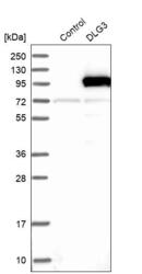
- Experimental details
- Western blot analysis in control (vector only transfected HEK293T lysate) and DLG3 over-expression lysate (Co-expressed with a C-terminal myc-DDK tag (~3.1 kDa) in mammalian HEK293T cells, LY402837).
- Sample type
- Human
- Protocol
- Protocol
