Explore
 Validate
 Learn
Learn Western blot
 ELISA
ELISA Immunocytochemistry
Antibody data
- Antibody Data
- Antigen structure
- References [3]
- Comments [0]
- Validations
- Immunocytochemistry [2]
- Immunohistochemistry [3]
- Other assay [4]
Submit
Validation data
Reference
Comment
Report error
- Product number
- 32-5800 - Provider product page

- Provider
- Invitrogen Antibodies
- Product name
- PTEN Monoclonal Antibody (2F4C9)
- Antibody type
- Monoclonal
- Antigen
- Recombinant full-length protein
- Description
- 32-5800 reacts specifically with the human PTEN protein. Reactivity was confirmed with cell lines which contain endogenous PTEN (NIH3T3, K652, and DU145 prostate cancer line) as well as transfected cell lines (Rat1 and 293T cells). This antibody detects the ~55 kDa PTEN protein and was successfully used in western blotting analysis of PTEN in breast cancer cell lines. No cross-reactivity to any other proteins has been detected.
- Reactivity
- Human, Mouse
- Host
- Mouse
- Isotype
- IgG
- Antibody clone number
- 2F4C9
- Vial size
- 100 μg
- Concentration
- 0.5 mg/mL
- Storage
- -20°C
Submitted references The ISG15-specific protease USP18 regulates stability of PTEN.
Epigenetic and genetic alterations of PTEN in hepatocellular carcinoma.
In vivo gene therapy of human bladder cancer with PTEN suppresses tumor growth, downregulates phosphorylated Akt, and increases sensitivity to doxorubicin.
Mustachio LM, Kawakami M, Lu Y, Rodriguez-Canales J, Mino B, Behrens C, Wistuba I, Bota-Rabassedas N, Yu J, Lee JJ, Roszik J, Zheng L, Liu X, Freemantle SJ, Dmitrovsky E
Oncotarget 2017 Jan 3;8(1):3-14
Oncotarget 2017 Jan 3;8(1):3-14
Epigenetic and genetic alterations of PTEN in hepatocellular carcinoma.
Wang L, Wang WL, Zhang Y, Guo SP, Zhang J, Li QL
Hepatology research : the official journal of the Japan Society of Hepatology 2007 May;37(5):389-96
Hepatology research : the official journal of the Japan Society of Hepatology 2007 May;37(5):389-96
In vivo gene therapy of human bladder cancer with PTEN suppresses tumor growth, downregulates phosphorylated Akt, and increases sensitivity to doxorubicin.
Tanaka M, Grossman HB
Gene therapy 2003 Sep;10(19):1636-42
Gene therapy 2003 Sep;10(19):1636-42
No comments: Submit comment
Supportive validation
- Submitted by
- Invitrogen Antibodies (provider)
- Main image
- Experimental details
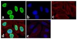
- Immunofluorescent analysis of PTEN was done on 70% confluent log phase HeLa cells. The cells were fixed with 4% paraformaldehyde for 15 minutes, permeabilized with 0.25% Triton X-100 for 10 minutes, and blocked with 5% BSA for 1 hour at room temperature. The cells were labeled with PTEN Mouse Monoclonal Antibody (Product # 32-5800) at 0.5 µg/mL in 1% BSA and incubated for 3 hours at room temperature and then labeled with Alexa Fluor 488 Rabbit Anti-Mouse IgG Secondary Antibody (Product # A-11059) at a dilution of 1:400 for 30 minutes at room temperature (Panel a: green). Nuclei (Panel b: blue) were stained with SlowFade Gold Antifade Mountant with DAPI (Product # S36938). F-actin (Panel c: red) was stained with Alexa Fluor 594 Phalloidin (Product # A12381). Panel d is a merged image showing nuclear localization and panel e is a no primary antibody control. The images were captured at 20X magnification.
- Submitted by
- Invitrogen Antibodies (provider)
- Main image

- Experimental details
- Immunofluorescent analysis of PTEN was done on 70% confluent log phase HeLa cells. The cells were fixed with 4% paraformaldehyde for 15 minutes, permeabilized with 0.25% Triton X-100 for 10 minutes, and blocked with 5% BSA for 1 hour at room temperature. The cells were labeled with PTEN Mouse Monoclonal Antibody (Product # 32-5800) at 0.5 µg/mL in 1% BSA and incubated for 3 hours at room temperature and then labeled with Alexa Fluor 488 Rabbit Anti-Mouse IgG Secondary Antibody (Product # A-11059) at a dilution of 1:400 for 30 minutes at room temperature (Panel a: green). Nuclei (Panel b: blue) were stained with SlowFade Gold Antifade Mountant with DAPI (Product # S36938). F-actin (Panel c: red) was stained with Alexa Fluor 594 Phalloidin (Product # A12381). Panel d is a merged image showing nuclear localization and panel e is a no primary antibody control. The images were captured at 20X magnification.

Supportive validation
- Submitted by
- Invitrogen Antibodies (provider)
- Main image
- Experimental details
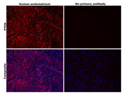
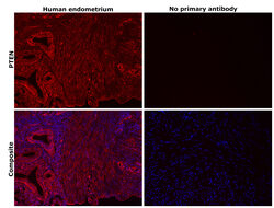
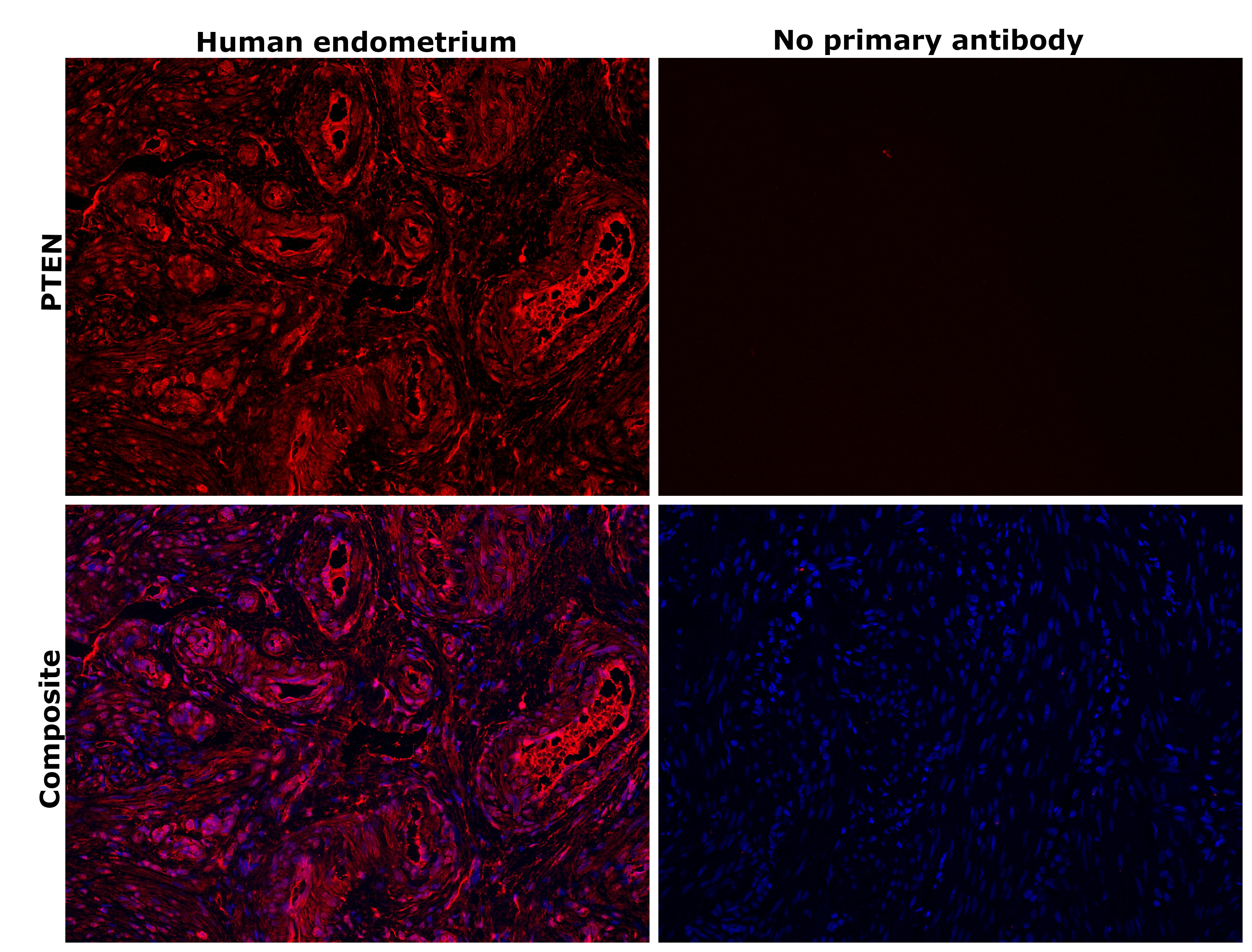
- Immunohistochemical analysis of PTEN was performed using formalin-fixed paraffin-embedded human endometrium tissue sections. To expose the target protein, heat-induced epitope retrieval was performed on de-paraffinized sections using eBioscience™ IHC Antigen Retrieval Solution - High pH (10X) (Product # 00-4956-58) diluted to 1X solution in water in a decloaking chamber at 110 degree Celsius for 15 minutes. Following antigen retrieval, the sections were blocked with 3% H2O2 for 1 hour at room temperature followed by 2% normal goat serum in 1X PBS for 45 minutes at room temperature and then probed with or without PTEN Monoclonal Antibody (2F4C9) (Product # 32-5800) at 1 µg/mL in 0.1% normal goat serum overnight at 4 degree Celsius in a humidified chamber. Detection was performed using Alexa Fluor™ 647 Tyramide SuperBoost™ Kit, goat anti-mouse IgG (Product # B40916). Nuclei were stained with DAPI (Product # D1306) and the sections were mounted using ProLong™ Glass Antifade Mountant (Product # P36984). The images were captured on EVOS™ M7000 Imaging System (Product # AMF7000) at 20X magnification and externally deconvoluted.

- Submitted by
- Invitrogen Antibodies (provider)
- Main image
- Experimental details
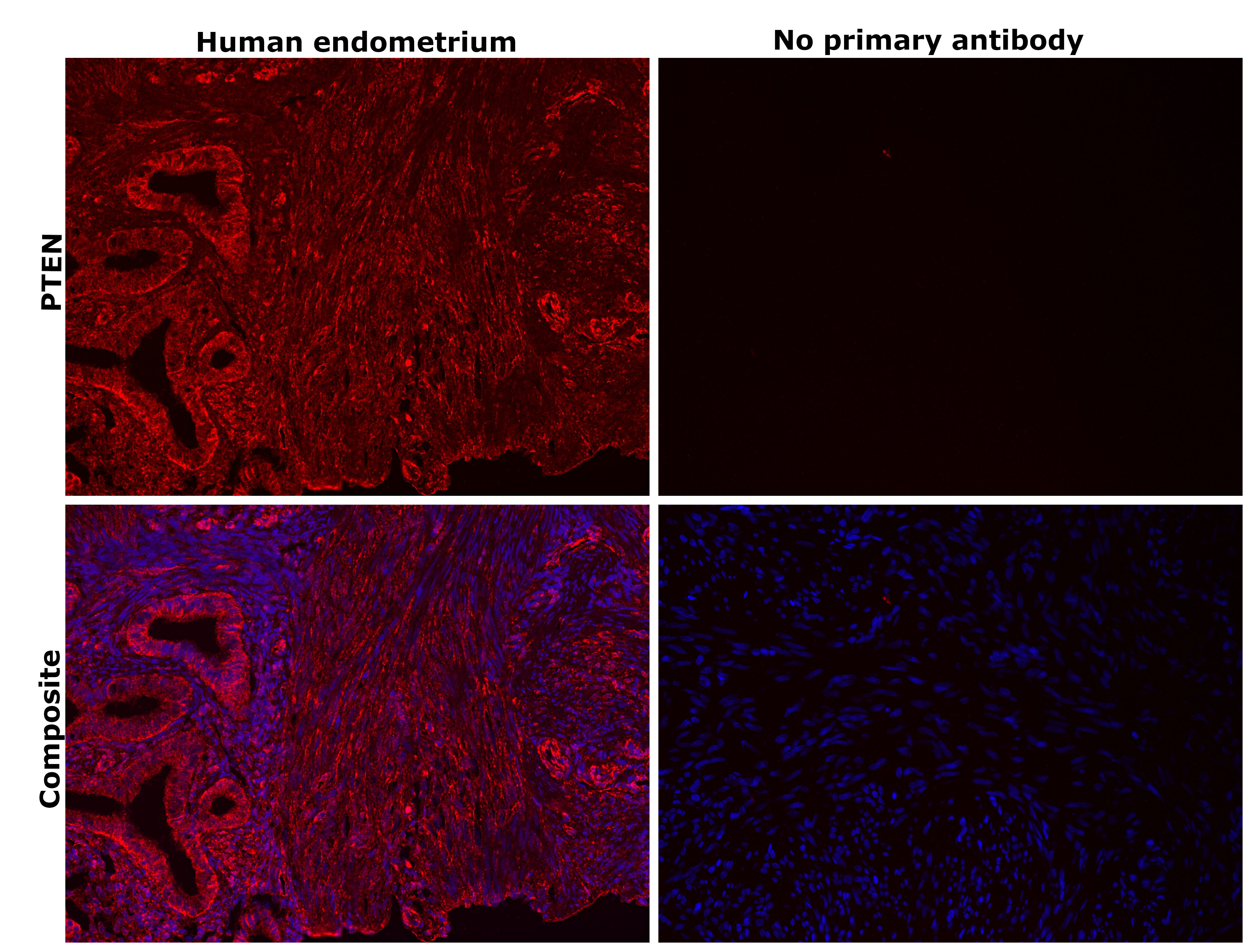
- Immunohistochemical analysis of PTEN was performed using formalin-fixed paraffin-embedded human endometrium tissue sections. To expose the target protein, heat-induced epitope retrieval was performed on de-paraffinized sections using eBioscience™ IHC Antigen Retrieval Solution - High pH (10X) (Product # 00-4956-58) diluted to 1X solution in water in a decloaking chamber at 110 degree Celsius for 15 minutes. Following antigen retrieval, the sections were blocked with 3% H2O2 for 1 hour at room temperature followed by 2% normal goat serum in 1X PBS for 45 minutes at room temperature and then probed with or without PTEN Monoclonal Antibody (2F4C9) (Product # 32-5800) at 1 µg/mL in 0.1% normal goat serum overnight at 4 degree Celsius in a humidified chamber. Detection was performed using Alexa Fluor™ 647 Tyramide SuperBoost™ Kit, goat anti-mouse IgG (Product # B40916). Nuclei were stained with DAPI (Product # D1306) and the sections were mounted using ProLong™ Glass Antifade Mountant (Product # P36984). The images were captured on EVOS™ M7000 Imaging System (Product # AMF7000) at 20X magnification and externally deconvoluted.
- Submitted by
- Invitrogen Antibodies (provider)
- Main image
- Experimental details
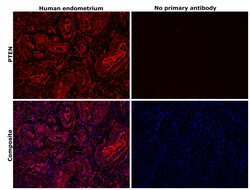
- Immunohistochemical analysis of PTEN was performed using formalin-fixed paraffin-embedded human endometrium tissue sections. To expose the target protein, heat-induced epitope retrieval was performed on de-paraffinized sections using eBioscience™ IHC Antigen Retrieval Solution - High pH (10X) (Product # 00-4956-58) diluted to 1X solution in water in a decloaking chamber at 110 degree Celsius for 15 minutes. Following antigen retrieval, the sections were blocked with 2% normal goat serum in 1X PBS for 45 minutes at room temperature and then probed with or without PTEN Monoclonal Antibody (2F4C9) (Product # 32-5800) at 5 µg/mL in 0.1% normal goat serum overnight at 4 degree Celsius in a humidified chamber. Detection was performed using Goat anti-Mouse IgG (H+L) Highly Cross-Adsorbed Secondary Antibody, Alexa Fluor™ Plus 647 (Product # A32728) at a dilution of 1:2,000 in 0.1% normal goat serum for 45 minutes at room temperature. ReadyProbes™ Tissue Autofluorescence Quenching Kit (Product # R37630) was used to quench autofluorescence from the tissues. Nuclei were stained with DAPI (Product # D1306) and the sections were mounted using ProLong™ Glass Antifade Mountant (Product # P36984). The images were captured on EVOS™ M7000 Imaging System (Product # AMF7000) at 20X magnification and externally deconvoluted.
Supportive validation
- Submitted by
- Invitrogen Antibodies (provider)
- Main image
- Experimental details
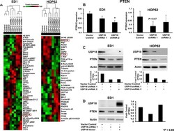
- Figure 1 RPPA-based protein profiling revealed USP18 regulated PTEN levels A. Independent RPPAs are displayed of ED1 and HOP62 lung cancer cell lines stably transfected with a control vector or one of two individual shRNAs against USP18. Signal intensities were normalized and transformed to linear values. RPPA highlighted species showing substantial differences in expression profiles between vector control and USP18 shRNA transfected lung cancer cells are represented by heat-maps for both ED1 and HOP62 lung cancer arrays. Red indicated high and green indicated low protein expression. Cell line clusters are shown as dendrograms. PTEN (red box) was highlighted as one of the proteins showing similar trends in both arrays. B. PTEN expression in the displayed ED1 and HOP62 transfected lung cancer cell lines was quantified using normalized linear values. Cell lines stably transfected with shRNAs against USP18 were compared relative to vector control transfectants. C. Representative immunoblots independently probed with USP18 and PTEN recognizing antibodies confirmed that engineered repression of USP18 decreased PTEN protein levels in ED1 and HOP62 lung cancer cells. PTEN expression was normalized to actin expression and compared between vector control and USP18 shRNA-transfected lung cancer cell lines. D. Decreased PTEN protein levels caused by loss of USP18 expression were rescued by transiently overexpressing GFP-tagged USP18 in ED1 lung cancer cells. Immunoblots were individuall

- Submitted by
- Invitrogen Antibodies (provider)
- Main image
- Experimental details
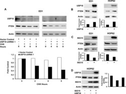
- Figure 2 Modifying USP18 and ISG15 protein levels altered PTEN protein stability A. Loss of USP18 expression decreased PTEN protein stability. ED1 murine lung cancer cells stably transfected with a vector control or with one of two individual shRNAs against USP18 were subjected to an 8 hour CHX treatment. PTEN expression was quantified relative to respective actin expression at indicated time points and normalized to the 0 hour (before CHX treatment) time point. B. Gain of USP18 expression increased PTEN levels. ED1 and HOP62 lung cancer cells were independently transfected with a USP18 expression plasmid or a control vector. PTEN expression was normalized to actin expression and compared between vector control and USP18 vector-transfected lung cancer cell lines. C. Engineered gain of ISG15 expression decreased PTEN protein levels. ED1 and HOP62 lung cancer cells were independently transfected with ISG15 or a vector control. PTEN was normalized to actin expression and compared between vector control and ISG15 vector-transfected lung cancer cell lines. D. Decreased PTEN levels conferred by overexpressed ISG15 were rescued by transiently overexpressing GFP-tagged USP18 in HOP62 lung cancer cells. Immunoblots were independently probed with GFP and PTEN recognizing antibodies. PTEN protein levels were normalized to actin expression and compared between vector control and ISG15 vector as well as ISG15 vector/USP18 vector-transfected lung cancer cell lines.

- Submitted by
- Invitrogen Antibodies (provider)
- Main image
- Experimental details
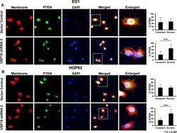
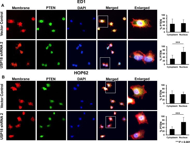
- Figure 3 Loss of USP18 expression decreased cytoplasmic PTEN protein A. ED1 and B. HOP62 lung cancer cell lines stably transfected with a vector control or shRNA against USP18 were fixed and stained for PTEN, sodium potassium ATPase, and DAPI. Cells were imaged by confocal microscopy and the magnification was 63X. Percent immunostaining of PTEN in the cytoplasm and nucleus was quantified and compared. Data are representative of 20 cells per group for each analyzed experiment.
- Submitted by
- Invitrogen Antibodies (provider)
- Main image

- Experimental details
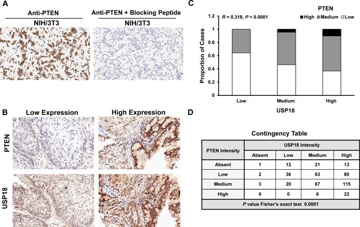
- Figure 5 PTEN and USP18 expression profiles are associated in human lung cancers A. Blocking peptide antagonized PTEN immunostaining in the NIH/3T3 (PTEN positive) cell line. B. Representative staining for human lung cancers with low PTEN and USP18 or high PTEN and USP18 expression profiles. All magnifications were 20X. C. PTEN and USP18 immunostaining are positively correlated in human lung cancer ( n = 461) and D. This association was confirmed using a contingency table by grouping negative, low, medium, or high PTEN and USP18 immunostaining profiles in lung cancer cases.