 Explore
 Validate
 Learn
Learn Western blot
 Immunohistochemistry
Antibody data
- Antibody Data
- Antigen structure
- References [1]
- Comments [0]
- Validations
- Immunohistochemistry [1]
- Flow cytometry [1]
- Other assay [1]
Submit
Validation data
Reference
Comment
Report error
- Product number
- MA5-14851 - Provider product page

- Provider
- Invitrogen Antibodies
- Product name
- PDGFRB Monoclonal Antibody (R.140.4)
- Antibody type
- Monoclonal
- Antigen
- Other
- Description
- It is not recommended to aliquot this antibody. This antibody is not cross-reactive with other PDGF receptor family members.
- Reactivity
- Human, Mouse, Rat
- Host
- Rabbit
- Isotype
- IgG
- Antibody clone number
- R.140.4
- Vial size
- 100 μL
- Concentration
- 7 μg/mL
- Storage
- -20°C
Submitted references The effects of focal adhesion kinase and platelet-derived growth factor receptor beta inhibition in a patient-derived xenograft model of primary and metastatic Wilms tumor.
Aye JM, Stafman LL, Williams AP, Garner EF, Stewart JE, Anderson JC, Mruthyunjayappa S, Waldrop MG, Goolsby CD, Markert HR, Quinn C, Marayati R, Mroczek-Musulman E, Willey CD, Yoon KJ, Whelan KF, Beierle EA
Oncotarget 2019 Sep 17;10(53):5534-5548
Oncotarget 2019 Sep 17;10(53):5534-5548
No comments: Submit comment
Supportive validation
- Submitted by
- Invitrogen Antibodies (provider)
- Main image

- Experimental details
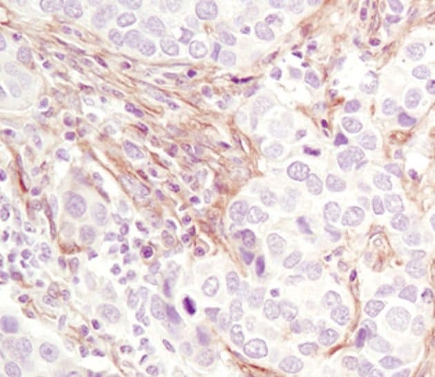
- Immunohistochemical analysis of PDGF Receptor beta in paraffin-embedded human lung carcinoma using a PDGF Receptor beta monoclonal antibody (Product # MA5-14851).
Supportive validation
- Submitted by
- Invitrogen Antibodies (provider)
- Main image
- Experimental details
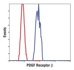
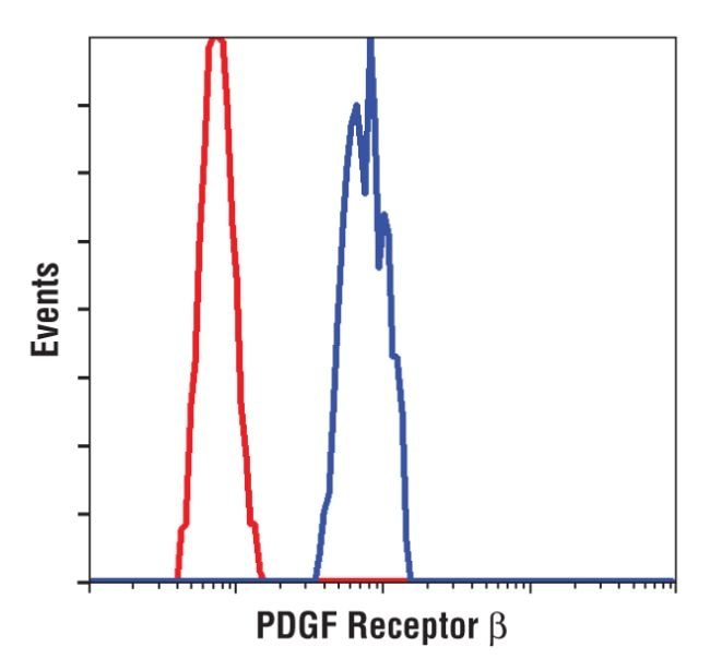
- Flow cytometric analysis of PDGF Receptor beta in NIH/3T3 cells using a PDGF Receptor beta monoclonal antibody (Product # MA5-14851) (blue) compared to a nonspecific negative control antibody (red).
Supportive validation
- Submitted by
- Invitrogen Antibodies (provider)
- Main image
- Experimental details
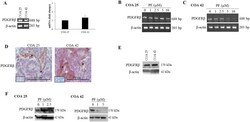
- Figure 4 PF-573,228 (PF) inhibition of FAK altered PDGFRbeta in COA 25 and COA 42 cells. ( A ) Real-time reverse transcription PCR and gel electrophoresis for PDGFRbeta were performed using 0.5 ug of extracted RNA from COA 25 and COA 42 cells. Abundance of PDGFRbeta mRNA did not significantly differ between COA 25 and COA 42 cells. ( B ) PCR for PDGFRbeta mRNA was performed using 0.5 ug of extracted RNA from COA 25 cells treated with increasing concentrations of PF. Abundance of mRNA for PDGFRbeta did not appear significantly different in PF-treated COA 25 cells. ( C ) PCR for PDGFRbeta mRNA was performed using 0.5 ug of extracted RNA from COA 42 cells treated with increasing concentrations of PF. Abundance of mRNA for PDGFRbeta was significantly decreased in PF-treated COA 42 cells. ( D ) Immunohistochemical staining with antibodies for PDGFRbeta was performed on PDXs COA 25 and COA 42. Negative controls were included for each sample ( inserts ). Staining for PDGFRbeta was positive and located in tumor cell cytoplasm and membrane ( dashed arrows ) and in tumor stroma ( solid arrows ) of PDXs COA 25 and COA 42. ( E ) Immunoblotting for PDGFRbeta was performed on COA 25 and COA 42 cell lysates. PDGFRbeta was detected in both PDXs. ( F ) COA 25 and COA 42 cells were treated for 24 hours with increasing concentrations of PF. Cell lysates were harvested and evaluated with immunoblotting for PDGFRbeta. Treatment with PF increased expression of PDGFRbeta in COA 25 cells while decre
