Explore
 Validate
 Learn
Learn Western blot
 Immunocytochemistry
 Immunoprecipitation
Antibody data
- Antibody Data
- Antigen structure
- References [3]
- Comments [0]
- Validations
- Immunocytochemistry [1]
Submit
Validation data
Reference
Comment
Report error
- Product number
- ALX-804-891-C100 - Provider product page

- Provider
- Enzo Life Sciences
- Product name
- AIM2 (human) monoclonal antibody (3B10)
- Antibody type
- Monoclonal
- Antigen
- Recombinant protein fragment
- Description
- Affinity purified.
- Reactivity
- Human
- Host
- Mouse
- Isotype
- IgG
- Antibody clone number
- 3B10
- Vial size
- 100 μg
- Storage
- 0
Submitted references AIM2 recognizes cytosolic dsDNA and forms a caspase-1-activating inflammasome with ASC.
AIM2 activates the inflammasome and cell death in response to cytoplasmic DNA.
Biochemical and growth regulatory activities of the HIN-200 family member and putative tumor suppressor protein, AIM2.
Hornung V, Ablasser A, Charrel-Dennis M, Bauernfeind F, Horvath G, Caffrey DR, Latz E, Fitzgerald KA
Nature 2009 Mar 26;458(7237):514-8
Nature 2009 Mar 26;458(7237):514-8
AIM2 activates the inflammasome and cell death in response to cytoplasmic DNA.
Fernandes-Alnemri T, Yu JW, Datta P, Wu J, Alnemri ES
Nature 2009 Mar 26;458(7237):509-13
Nature 2009 Mar 26;458(7237):509-13
Biochemical and growth regulatory activities of the HIN-200 family member and putative tumor suppressor protein, AIM2.
Cresswell KS, Clarke CJ, Jackson JT, Darcy PK, Trapani JA, Johnstone RW
Biochemical and biophysical research communications 2005 Jan 14;326(2):417-24
Biochemical and biophysical research communications 2005 Jan 14;326(2):417-24
No comments: Submit comment
Supportive validation
- Submitted by
- Enzo Life Sciences (provider)
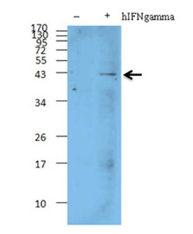
- Main image
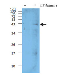
- Experimental details
- Western blot analyisis of AIM2, mAb (c3B10). HL-60 cells were treated with human IFN_ recombinant protein (570202) at 10 ng/ml for 16 hours. The untreated cells were used as the negative control. Cell lysates from control cells and the hIFN_ treated cells were resolved by electrophoresis, transferred to nitrocellulose, and probed with AIM2, mAb (c3B10). Proteins were visualized using a goat anti-mouse secondary conjugated to HRP and a chemiluminescence detection system.