 Explore
 Validate
 Learn
LearnNBP2-23669
antibody from Novus Biologicals
Targeting: GPBAR1
BG37, GPCR, GPCR19, GPR131, M-BAR, MGC40597, TGR5
 Western blot
 Immunocytochemistry
Antibody data
- Antibody Data
- Antigen structure
- References [1]
- Comments [0]
- Validations
- Western blot [1]
- Immunohistochemistry [1]
Submit
Validation data
Reference
Comment
Report error
- Product number
- NBP2-23669 - Provider product page

- Provider
- Novus Biologicals
- Product name
- Rabbit Polyclonal TGR5/GPBAR1 Antibody
- Antibody type
- Polyclonal
- Description
- Immunogen affinity purified.
- Reactivity
- Human, Mouse, Rat
- Host
- Rabbit
- Isotype
- IgG
- Vial size
- 0.1 ml
- Concentration
- 1.0 mg/ml
- Storage
- Store at -20C. Avoid freeze-thaw cycles.
Submitted references Lithocholic acid, a bacterial metabolite reduces breast cancer cell proliferation and aggressiveness.
Mikó E, Vida A, Kovács T, Ujlaki G, Trencsényi G, Márton J, Sári Z, Kovács P, Boratkó A, Hujber Z, Csonka T, Antal-Szalmás P, Watanabe M, Gombos I, Csoka B, Kiss B, Vígh L, Szabó J, Méhes G, Sebestyén A, Goedert JJ, Bai P
Biochimica et biophysica acta. Bioenergetics 2018 Sep;1859(9):958-974
Biochimica et biophysica acta. Bioenergetics 2018 Sep;1859(9):958-974
No comments: Submit comment
Supportive validation
- Submitted by
- Novus Biologicals (provider)
- Main image
- Experimental details
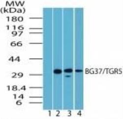
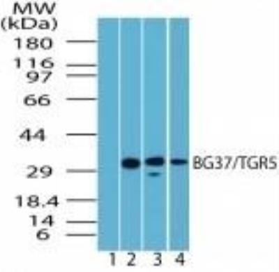
- Western Blot: TGR5/GPBAR1 Antibody [NBP2-23669] - Analysis of spleen lysate. (1) Pre-immune sera. TGR5/GPBAR1 antibody tested on (2) Human 1:1000, (3) Mouse 1:5000, and (4) Rat 1:5000 spleen lysates.
Supportive validation
- Submitted by
- Novus Biologicals (provider)
- Main image
- Experimental details
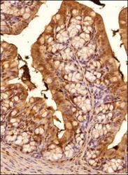
- Immunohistochemistry-Paraffin: TGR5/GPBAR1 Antibody [NBP2-23669] - Tissue section of mouse colon using TGR5/GPBAR1 antibody at 1:200 dilution. The signal was detected using HRP-labelled secondary antibody and DAB staining which followed counterstaining with hematoxylin. This TGR5/GPBAR1 antibody generated a very specific a cytoplasmic staining primarily in the columner epithelial cells. A subset of cells from mucosa muscularis and the lamina propria also showed TGR5 positivity. Some cells in all histological layers depicted a nuclear staining also.
