Explore
 Validate
 Learn
LearnMA5-17266
antibody from Invitrogen Antibodies
Targeting: PEX19
D1S2223E, HK33, PMP1, PMPI, PXF, PXMP1
 Western blot
 Immunocytochemistry
Antibody data
- Antibody Data
- Antigen structure
- References [2]
- Comments [0]
- Validations
- Immunocytochemistry [2]
- Flow cytometry [2]
- Other assay [2]
Submit
Validation data
Reference
Comment
Report error
- Product number
- MA5-17266 - Provider product page

- Provider
- Invitrogen Antibodies
- Product name
- PEX19 Monoclonal Antibody (GT554)
- Antibody type
- Monoclonal
- Antigen
- Recombinant full-length protein
- Description
- Recommended positive controls: Jurkat, Raji, K562, THP-1, HL-60, NCI-H929, mouse liver. Predicted reactivity: Mouse (91%), Rat (93%), Bovine (95%). Store product as a concentrated solution. Centrifuge briefly prior to opening the vial.
- Reactivity
- Human, Mouse
- Host
- Mouse
- Isotype
- IgG
- Antibody clone number
- GT554
- Vial size
- 100 μL
- Concentration
- 1 mg/mL
- Storage
- Store at 4°C short term. For long term storage, store at -20°C, avoiding freeze/thaw cycles.
Submitted references Integrative Informatics Analysis of Transcriptome and Identification of Interacted Genes in the Glomeruli and Tubules in CKD.
Allosteric modulation of peroxisomal membrane protein recognition by farnesylation of the peroxisomal import receptor PEX19.
Liu L, Ma F, Hao Y, Yi Z, Yu X, Xu B, Wei C, Hu J
Frontiers in medicine 2020;7:615306
Frontiers in medicine 2020;7:615306
Allosteric modulation of peroxisomal membrane protein recognition by farnesylation of the peroxisomal import receptor PEX19.
Emmanouilidis L, Schütz U, Tripsianes K, Madl T, Radke J, Rucktäschel R, Wilmanns M, Schliebs W, Erdmann R, Sattler M
Nature communications 2017 Mar 10;8:14635
Nature communications 2017 Mar 10;8:14635
No comments: Submit comment
Supportive validation
- Submitted by
- Invitrogen Antibodies (provider)
- Main image

- Experimental details
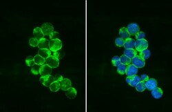
- Immunocytochemistry-Immunofluorescence analysis of PEX19 was performed in Jurkat cells fixed in 4% paraformaldehyde at RT for 15 min. Green: PEX19 Monoclonal Antibody (GT554) (Product # MA5-17266) diluted at 1:500. Blue: Fluoroshield with DAPI.

- Submitted by
- Invitrogen Antibodies (provider)
- Main image
- Experimental details
- Immunocytochemistry-Immunofluorescence analysis of PEX19 was performed in Jurkat cells fixed in 4% paraformaldehyde at RT for 15 min. Green: PEX19 Monoclonal Antibody (GT554) (Product # MA5-17266) diluted at 1:500. Blue: Fluoroshield with DAPI.

Supportive validation
- Submitted by
- Invitrogen Antibodies (provider)
- Main image

- Experimental details
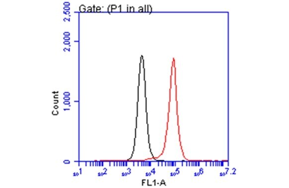
- Flow Cytometry analysis of ABCA1 was performed in THP-1 cells using ABCA1 Polyclonal Antibody (Product # PA1-32129) (red) at a dilution of 1:50. Black: Unlabelled sample was used as a control. Acquisition of 20,000 events were collected using a Dylight 488-conjugated secondary antibody for FACS analysis.

- Submitted by
- Invitrogen Antibodies (provider)
- Main image

- Experimental details
- Flow Cytometry analysis of ABCA1 was performed in THP-1 cells using ABCA1 Polyclonal Antibody (Product # PA1-32129) (red) at a dilution of 1:50. Black: Unlabelled sample was used as a control. Acquisition of 20,000 events were collected using a Dylight 488-conjugated secondary antibody for FACS analysis.
Supportive validation
- Submitted by
- Invitrogen Antibodies (provider)
- Main image
- Experimental details
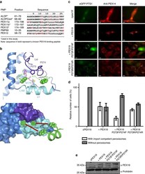
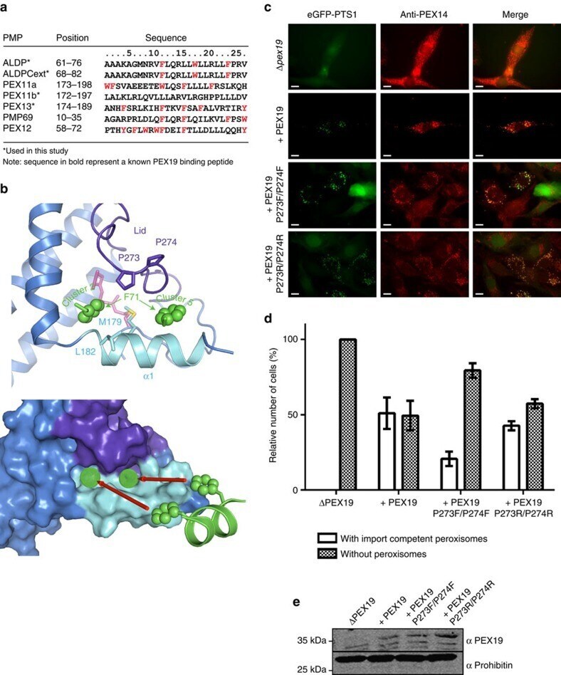
- Figure 5 Recognition of a PMP-derived peptide by the farnesylated PEX19 CTD. ( a ) Amino acid sequences of peroxisomal membrane proteins. Aromatic residues are highlighted in red; regions known to be involved in PEX19 binding are shown in bold. ( b ) Docking models showing two binding pockets for aromatic side chains of the PMP-derived peptide (green stick and ball representation). Top: The PEX19 lid and helix alpha1 are coloured dark blue and cyan, respectively. Side chains of M179, Leu182, Pro272 and Pro273 are shown as sticks. Bottom: Surface representation of the PEX19 CTD indicating the hydrophobic cavities that accommodate the aromatic side chain of Phe71 in the ALDP peptide. ( c ) In vivo effects of mutations of the PMP binding site of PEX19. Immunofluorescence microscopy images of PEX19-deficient human fibroblasts transfected with bicistronic vectors encoding for eGFP-PTS1 and different PEX19 variants. The same plasmid lacking PEX19 was used as a negative control (DeltaPEX19) showing diffuse staining due to mitochondrial mislocalization for both peroxisomal marker proteins, eGFP-PTS1 (matrix protein) and PEX14 (PMP). The same plasmid lacking PEX19 was used as a negative control (DeltaPEX19) showing cytosolic and mitochondrial mislocalization for the peroxisomal marker proteins eGFP-PTS1 (matrix protein) and PEX14 (PMP), respectively. A congruent punctate pattern of eGFP-SKL and PEX14 indicates formation of import-competent peroxisomes 72 h after transfection. Scale ba
- Submitted by
- Invitrogen Antibodies (provider)
- Main image
- Experimental details
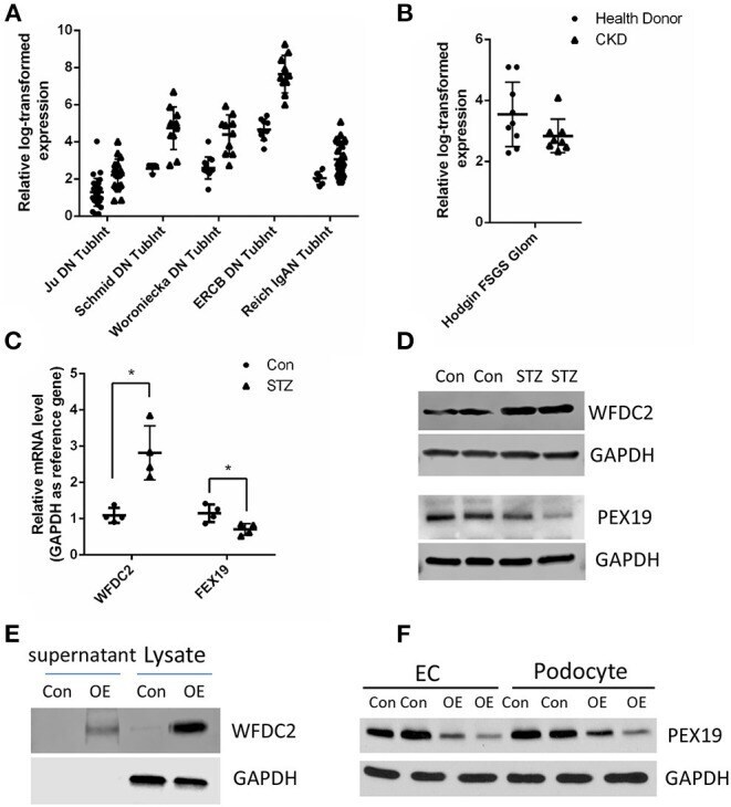
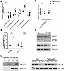
- Figure 5 Verification of WFDC2 and PEX19 interaction in HK2, podocyte, and glomerular endothelial cells. (A) Relative WFDC2 transcript levels were upregulated in many diabetic nephropathy human and mice datasets in the kidney tubule. (B) Relative FEX19 transcript levels were downregulated in FSGS in the kidney glomeruli in Nephroseq database. (C,D) WFDC2 in the tubule was upregulated, and FEX19 in the glomeruli was downregulated in STZ-induced diabetic mice by qPCR and western blot. (E) Western blot shows WFDC2 overexpression in HK2 cell. (F) PEX19 protein level was decreased with WFDC2 treatment in podocyte and glomerular endothelial cells by western blot. Con, WFDC2 empty vector; OE, WFDC2 overexpression vector; EC, glomerular endothelial cells. (A-C) Values are mean +- SEM; * p