 Explore
 Validate
 Learn
LearnPA5-22129
antibody from Invitrogen Antibodies
Targeting: PEX19
D1S2223E, HK33, PMP1, PMPI, PXF, PXMP1
 Western blot
 Immunocytochemistry
Antibody data
- Antibody Data
- Antigen structure
- References [3]
- Comments [0]
- Validations
- Immunocytochemistry [5]
- Immunohistochemistry [2]
- Other assay [2]
Submit
Validation data
Reference
Comment
Report error
- Product number
- PA5-22129 - Provider product page

- Provider
- Invitrogen Antibodies
- Product name
- PEX19 Polyclonal Antibody
- Antibody type
- Polyclonal
- Antigen
- Recombinant full-length protein
- Description
- Recommended positive controls: Jurkat, Raji, NCI-H929, K562, THP-1, HL-60, mouse liver, PC-12, Rat-2. Predicted reactivity: Mouse (91%), Rat (93%), Bovine (95%). Store product as a concentrated solution. Centrifuge briefly prior to opening the vial.
- Reactivity
- Human, Mouse, Rat
- Host
- Rabbit
- Isotype
- IgG
- Vial size
- 100 μL
- Concentration
- 1.01 mg/mL
- Storage
- Store at 4°C short term. For long term storage, store at -20°C, avoiding freeze/thaw cycles.
Submitted references Dual role of USP30 in controlling basal pexophagy and mitophagy.
The peroxisome biogenesis factors posttranslationally target reticulon homology domain-containing proteins to the endoplasmic reticulum membrane.
Parkinson's disease-associated mutant VPS35 causes mitochondrial dysfunction by recycling DLP1 complexes.
Marcassa E, Kallinos A, Jardine J, Rusilowicz-Jones EV, Martinez A, Kuehl S, Islinger M, Clague MJ, Urbé S
EMBO reports 2018 Jul;19(7)
EMBO reports 2018 Jul;19(7)
The peroxisome biogenesis factors posttranslationally target reticulon homology domain-containing proteins to the endoplasmic reticulum membrane.
Yamamoto Y, Sakisaka T
Scientific reports 2018 Feb 2;8(1):2322
Scientific reports 2018 Feb 2;8(1):2322
Parkinson's disease-associated mutant VPS35 causes mitochondrial dysfunction by recycling DLP1 complexes.
Wang W, Wang X, Fujioka H, Hoppel C, Whone AL, Caldwell MA, Cullen PJ, Liu J, Zhu X
Nature medicine 2016 Jan;22(1):54-63
Nature medicine 2016 Jan;22(1):54-63
No comments: Submit comment
Supportive validation
- Submitted by
- Invitrogen Antibodies (provider)
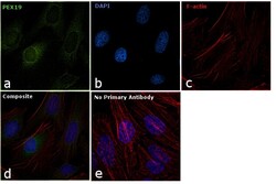
- Main image

- Experimental details
- Immunofluorescent analysis of PEX19 in methanol-fixed HeLa cells using a PEX19 polyclonal antibody (Product # PA5-22129) (Green) at a 1:500 dilution. Alpha-tubulin filaments were labeled with Product # PA5-29281 (Red) at a 1:2000.
- Submitted by
- Invitrogen Antibodies (provider)
- Main image

- Experimental details
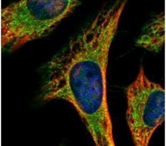
- Confocal immunofluorescence analysis (Olympus FV10i) of methanol-fixed HeLa, using PEX19 antibody (Product # PA5-22129) (Green) at 1:500 dilution. Alpha-tubulin filaments were labeled with (Product # MA1-25054) (Red) at 1:2,000.

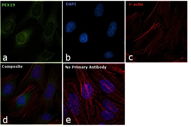
- Submitted by
- Invitrogen Antibodies (provider)
- Main image

- Experimental details
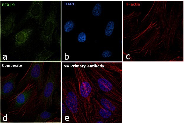
- Immunofluorescence analysis of PEX19 was performed using 70% confluent log phase HeLa cells. The cells were fixed with 4% paraformaldehyde for 10 minutes, permeabilized with 0.1% Triton™ X-100 for 10 minutes, and blocked with 1% BSA for 1 hour at room temperature. The cells were labeled with PEX19 Rabbit Polyclonal Antibody (Product # PA5-22129) at 5 µg/mL in 0.1% BSA and incubated overnight at 4 degree and then labeled with Goat anti-Rabbit IgG (H+L) Superclonal™ Secondary Antibody, Alexa Fluor® 488 conjugate (Product # A27034) at a dilution of 1:2000 for 45 minutes at room temperature (Panel a: green). Nuclei (Panel b: blue) were stained with SlowFade® Gold Antifade Mountant with DAPI (Product # S36938). F-actin (Panel c: red) was stained with Rhodamine Phalloidin (Product # R415, 1:300). Panel d represents the merged image showing cytoplasmic localization. Panel e represents control cells with no primary antibody to assess background. The images were captured at 60X magnification.
- Submitted by
- Invitrogen Antibodies (provider)
- Main image
- Experimental details
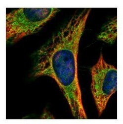
- Confocal immunofluorescence analysis (Olympus FV10i) of methanol-fixed HeLa, using PEX19 antibody (Product # PA5-22129) (Green) at 1:500 dilution. Alpha-tubulin filaments were labeled with (Product # MA1-25054) (Red) at 1:2,000.
- Submitted by
- Invitrogen Antibodies (provider)
- Main image
- Experimental details
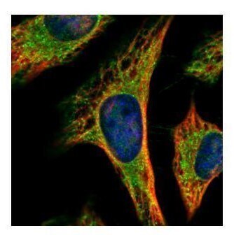
- Immunofluorescence analysis of PEX19 was performed using 70% confluent log phase HeLa cells. The cells were fixed with 4% paraformaldehyde for 10 minutes, permeabilized with 0.1% Triton™ X-100 for 10 minutes, and blocked with 1% BSA for 1 hour at room temperature. The cells were labeled with PEX19 Rabbit Polyclonal Antibody (Product # PA5-22129) at 5 µg/mL in 0.1% BSA and incubated overnight at 4 degree and then labeled with Goat anti-Rabbit IgG (Heavy Chain) Superclonal™ Secondary Antibody, Alexa Fluor® 488 conjugate (Product # A27034) at a dilution of 1:2000 for 45 minutes at room temperature (Panel a: green). Nuclei (Panel b: blue) were stained with SlowFade® Gold Antifade Mountant with DAPI (Product # S36938). F-actin (Panel c: red) was stained with Rhodamine Phalloidin (Product # R415, 1:300). Panel d represents the merged image showing cytoplasmic localization. Panel e represents control cells with no primary antibody to assess background. The images were captured at 60X magnification.
Supportive validation
- Submitted by
- Invitrogen Antibodies (provider)
- Main image
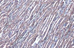
- Experimental details
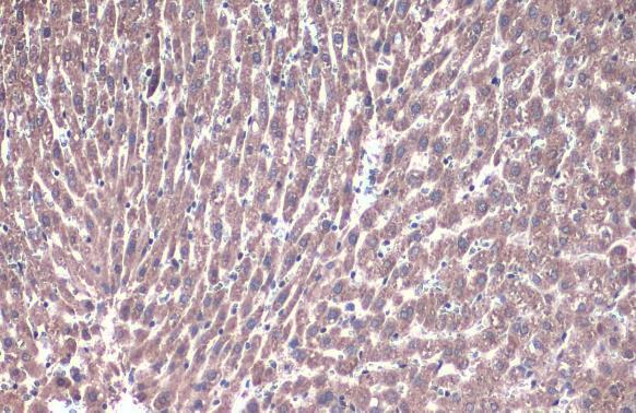
- PEX19 Polyclonal Antibody detects PEX19 protein at cytoplasm by immunohistochemical analysis. Sample: Paraffin-embedded mouse kidney. PEX19 stained by PEX19 Polyclonal Antibody (Product # PA5-22129) diluted at 1:1,000. Antigen Retrieval: Citrate buffer, pH 6.0, 15 min.
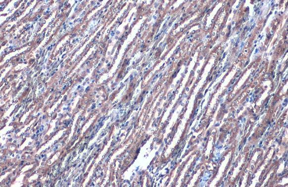
- Submitted by
- Invitrogen Antibodies (provider)
- Main image
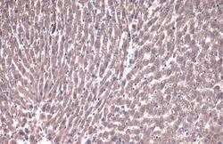
- Experimental details
- PEX19 Polyclonal Antibody detects PEX19 protein at cytoplasm by immunohistochemical analysis. Sample: Paraffin-embedded rat liver. PEX19 stained by PEX19 Polyclonal Antibody (Product # PA5-22129) diluted at 1:1,000. Antigen Retrieval: Citrate buffer, pH 6.0, 15 min.
Supportive validation
- Submitted by
- Invitrogen Antibodies (provider)
- Main image
- Experimental details
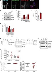
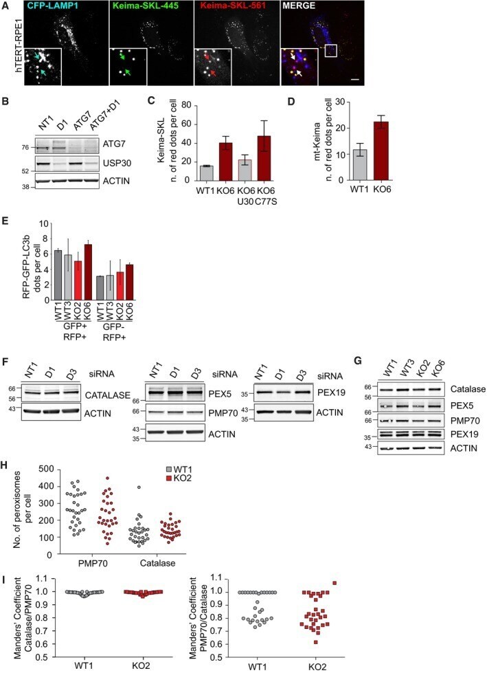
- Figure EV3 Basal autophagic flux and peroxisome abundance are not affected in USP30 KO cells Representative images of hTERT-RPE1 cells transfected with Keima-SKL and CFP-LAMP1 for 48 h, fixed and analysed by 3i-spinning disk confocal microscopy. Scale bar: 10 mum. Arrows indicate Keima-SKL ""red"" puncta that colocalise with the lysosomal marker CFP-LAMP1. Representative Western blot of cells analysed in Fig 5 B. Keima-SKL ""red"" puncta in USP30 KO or WT hTERT-RPE1 cells. Keima-SKL was either transfected on its own or together with USP30-GFP and USP30C77S-GFP for 48 h. Two independent experiments were analysed, 20 cells per experiment, mean +- range. Quantification of the percentage of mt-Keima ""red"" puncta in USP30 KO6 or WT1 hTERT-RPE1 cells ( n = 2 independent experiments, 20 cells per experiment, mean +- range). hTERT-RPE1 USP30 WT and KO cells (WT1, WT3, KO2 and KO6) were transfected with RFP-GFP-LC3b for 48 h prior to imaging by 3i-spinning disk confocal microscopy. Shown are representative images and the numbers of RFP-GFP-LC3b puncta per cell as an assessment of basal autophagic flux. Two independent experiments were analysed, 20 cells per experiment, mean +- range. hTERT-RPE1 cells were transfected with non-targeting (NT1) or USP30 targeting siRNA (D1 and D3) for 72 h and lysed in RIPA buffer. Protein samples were analysed by SDS-PAGE and Western blots probed for catalase, PMP70, PEX5 and PEX19. hTERT-RPE1 USP30 WT and KO cells (WT1, WT3, KO2 and KO6) were lysed i
- Submitted by
- Invitrogen Antibodies (provider)
- Main image
- Experimental details
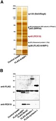
- Figure 2 Identification of PEX19 as a protein that selectively binds to the nascent RHD-containing proteins. ( A ) Purification of Arl6IP1-binding proteins from the rabbit reticulocyte lysates. The rabbit reticulocyte lysate synthesizing FLAG-Arl6IP1 was incubated with the anti-FLAG mAb-immobilized agarose. To check the contamination, the rabbit reticulocyte lysate without synthesis of FLAG-Arl6IP1 (control lysate) was also incubated with the anti-FLAG mAb-immobilized agarose. The bound proteins were eluted by competition with a large excess of free FLAG peptide and subjected to SDS-PAGE followed by silver staining. p120, p60, p55, p40 and p29 were identified as Bat3 (also known as Bag6), proteasome 26 S subunit ATPase1, SRP54, PEX19 and ribosomal protein L10a, respectively, by mass spectrometry. ( B ) Selective binding of PEX19 to the nascent RHD-containing proteins. The rabbit reticulocyte lysates synthesizing FLAG-Arl6IP1, FLAG-Rtn4C, FLAG-atlastin-1 and FLAG-syntaxin-1 were supplemented with 1 mM puromycin, followed by incubation with anti-FLAG mAb-immobilized agarose. The bound proteins were eluted with the SDS sample buffer with boiling and subjected to SDS-PAGE followed by Western blotting with the anti-PEX19 pAb and the anti-FLAG pAb.
