Explore
 Validate
 Learn
Learn Western blot
 Immunocytochemistry
Antibody data
- Antibody Data
- Antigen structure
- References [5]
- Comments [0]
- Validations
- Immunocytochemistry [1]
Submit
Validation data
Reference
Comment
Report error
- Product number
- MAB3491-100 - Provider product page

- Provider
- R&D Systems
- Product name
- Human VEGFR3/Flt-4 Antibody
- Antibody type
- Monoclonal
- Description
- Protein A or G purified from hybridoma culture supernatant. Detects human VEGFR3/Flt-4 in direct ELISAs and Western blots. In direct ELISAs and Western blots, approximately 25-30% cross-reactivity with recombinant mouse VEGFR3 is observed and no cross-reactivity with recombinant human (rh) VEGFR1 or rhVEGFR2 is observed.
- Reactivity
- Human
- Host
- Mouse
- Conjugate
- Unconjugated
- Antigen sequence
P35916- Isotype
- IgG
- Antibody clone number
- 54703
- Vial size
- 100 ug
- Storage
- Use a manual defrost freezer and avoid repeated freeze-thaw cycles. 12 months from date of receipt, -20 to -70 °C as supplied. 1 month, 2 to 8 °C under sterile conditions after reconstitution. 6 months, -20 to -70 °C under sterile conditions after reconstitution.
Submitted references Correlation of Vascular Endothelial Growth Factor subtypes and their receptors with melanoma progression: A next-generation Tissue Microarray (ngTMA) automated analysis.
Heparins that block VEGF-A-mediated von Willebrand factor fiber generation are potent inhibitors of hematogenous but not lymphatic metastasis.
Further evidence for expression and function of the VEGF-C/VEGFR-3 axis in cancer cells.
Vascular endothelial growth factor can signal through platelet-derived growth factor receptors.
The VEGF-C/Flt-4 axis promotes invasion and metastasis of cancer cells.
Seyed Jafari SM, Wiedmer C, Cazzaniga S, Frangež Ž, Shafighi M, Beltraminelli H, Weber B, Simon HU, Hunger RE
PloS one 2018;13(11):e0207019
PloS one 2018;13(11):e0207019
Heparins that block VEGF-A-mediated von Willebrand factor fiber generation are potent inhibitors of hematogenous but not lymphatic metastasis.
Goertz L, Schneider SW, Desch A, Mayer FT, Koett J, Nowak K, Karampinis I, Bohlmann MK, Umansky V, Bauer AT
Oncotarget 2016 Oct 18;7(42):68527-68545
Oncotarget 2016 Oct 18;7(42):68527-68545
Further evidence for expression and function of the VEGF-C/VEGFR-3 axis in cancer cells.
Su JL, Chen PS, Chien MH, Chen PB, Chen YH, Lai CC, Hung MC, Kuo ML
Cancer cell 2008 Jun;13(6):557-60
Cancer cell 2008 Jun;13(6):557-60
Vascular endothelial growth factor can signal through platelet-derived growth factor receptors.
Ball SG, Shuttleworth CA, Kielty CM
The Journal of cell biology 2007 May 7;177(3):489-500
The Journal of cell biology 2007 May 7;177(3):489-500
The VEGF-C/Flt-4 axis promotes invasion and metastasis of cancer cells.
Su JL, Yang PC, Shih JY, Yang CY, Wei LH, Hsieh CY, Chou CH, Jeng YM, Wang MY, Chang KJ, Hung MC, Kuo ML
Cancer cell 2006 Mar;9(3):209-23
Cancer cell 2006 Mar;9(3):209-23
No comments: Submit comment
Supportive validation
- Submitted by
- R&D Systems (provider)
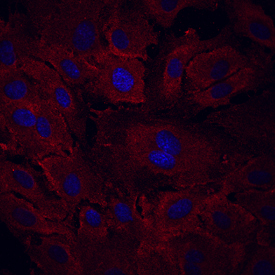
- Main image
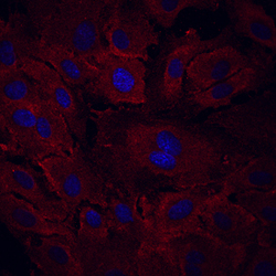
- Experimental details
- VEGFR3/Flt-4 in HUVEC Human Cells. VEGFR3/Flt-4 was detected in immersion fixed HUVEC human umbilical vein endothelial cells using Mouse Anti-Human VEGFR3/Flt-4 Monoclonal Antibody (Catalog # MAB3491) at 10 µg/mL for 3 hours at room temperature. Cells were stained using the NorthernLights™ 557-conjugated Anti-Mouse IgG Secondary Antibody (red; Catalog # NL007) and counterstained with DAPI (blue). Specific staining was localized to cell surfaces and cytoplasm. View our protocol for Fluorescent ICC Staining of Cells on Coverslips.