Explore
 Validate
 Learn
Learn Western blot
 Immunohistochemistry
Antibody data
- Antibody Data
- Antigen structure
- References [1]
- Comments [0]
- Validations
- Immunohistochemistry [4]
- Other assay [1]
Submit
Validation data
Reference
Comment
Report error
- Product number
- PA5-16871 - Provider product page

- Provider
- Invitrogen Antibodies
- Product name
- VEGF Receptor 3 Polyclonal Antibody
- Antibody type
- Polyclonal
- Antigen
- Synthetic peptide
- Description
- PA5-16871 targets Flt-4 in immunohistochemistry (paraffin), immunoprecipitation, and Western blot applications and shows reactivity with human, mouse, and rat samples. The PA5-16871 immunogen is a synthetic peptide derived from C-terminus of human Flt-4.
- Reactivity
- Human, Rat
- Host
- Rabbit
- Isotype
- IgG
- Vial size
- 500 μL
- Concentration
- Conc. Not Determined
- Storage
- -20°C, Avoid Freeze/Thaw Cycles
Submitted references Combined mTOR and MEK inhibition is an effective therapy in a novel mouse model for angiosarcoma.
Chadwick ML, Lane A, Thomas D, Smith AR, White AR, Davidson D, Feng Y, Boscolo E, Zheng Y, Adams DM, Gupta A, Veillette A, Chow LML
Oncotarget 2018 May 15;9(37):24750-24765
Oncotarget 2018 May 15;9(37):24750-24765
No comments: Submit comment
Supportive validation
- Submitted by
- Invitrogen Antibodies (provider)
- Main image

- Experimental details
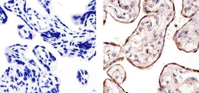
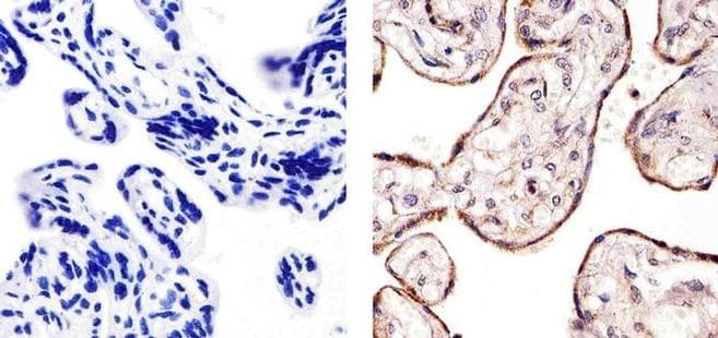
- Immunohistochemistry analysis of Flt-4 showing staining in the membrane of paraffin-embedded human placenta tissue (right) compared to a negative control without primary antibody (left). To expose target proteins, antigen retrieval was performed using 10mM sodium citrate (pH 6.0), microwaved for 8-15 min. Following antigen retrieval, tissues were blocked in 3% H2O2-methanol for 15 min at room temperature, washed with ddH2O and PBS, and then probed with a Flt-4 Rabbit Polyclonal Antibody (Product # PA5-16871) diluted in 3% BSA-PBS at a dilution of 1:20 for 1 hour at 37°C in a humidified chamber. Tissues were washed extensively in PBST and detection was performed using an HRP-conjugated secondary antibody followed by colorimetric detection using a DAB kit. Tissues were counterstained with hematoxylin and dehydrated with ethanol and xylene to prep for mounting.
- Submitted by
- Invitrogen Antibodies (provider)
- Main image
- Experimental details
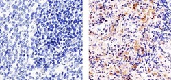
- Immunohistochemistry analysis of Flt-4 showing staining in the cytoplasm and membrane of paraffin-embedded mouse spleen tissue (right) compared to a negative control without primary antibody (left). To expose target proteins, antigen retrieval was performed using 10mM sodium citrate (pH 6.0), microwaved for 8-15 min. Following antigen retrieval, tissues were blocked in 3% H2O2-methanol for 15 min at room temperature, washed with ddH2O and PBS, and then probed with a Flt-4 Rabbit Polyclonal Antibody (Product # PA5-16871) diluted in 3% BSA-PBS at a dilution of 1:20 for 1 hour at 37°C in a humidified chamber. Tissues were washed extensively in PBST and detection was performed using an HRP-conjugated secondary antibody followed by colorimetric detection using a DAB kit. Tissues were counterstained with hematoxylin and dehydrated with ethanol and xylene to prep for mounting.

- Submitted by
- Invitrogen Antibodies (provider)
- Main image
- Experimental details
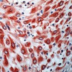
- Formalin-fixed, paraffin-embedded human placenta stained with anti-flt-4, . using peroxidase conjugate and AEC chromogen. Note cytoplasmic staining of trophoblasts.

- Submitted by
- Invitrogen Antibodies (provider)
- Main image

- Experimental details
- Immunohistochemistry analysis of Flt-4 showing staining in the membrane of paraffin-embedded human placenta tissue (right) compared to a negative control without primary antibody (left). To expose target proteins, antigen retrieval was performed using 10mM sodium citrate (pH 6.0), microwaved for 8-15 min. Following antigen retrieval, tissues were blocked in 3% H2O2-methanol for 15 min at room temperature, washed with ddH2O and PBS, and then probed with a Flt-4 Rabbit Polyclonal Antibody (Product # PA5-16871) diluted in 3% BSA-PBS at a dilution of 1:20 for 1 hour at 37°C in a humidified chamber. Tissues were washed extensively in PBST and detection was performed using an HRP-conjugated secondary antibody followed by colorimetric detection using a DAB kit. Tissues were counterstained with hematoxylin and dehydrated with ethanol and xylene to prep for mounting.
Supportive validation
- Submitted by
- Invitrogen Antibodies (provider)
- Main image
- Experimental details
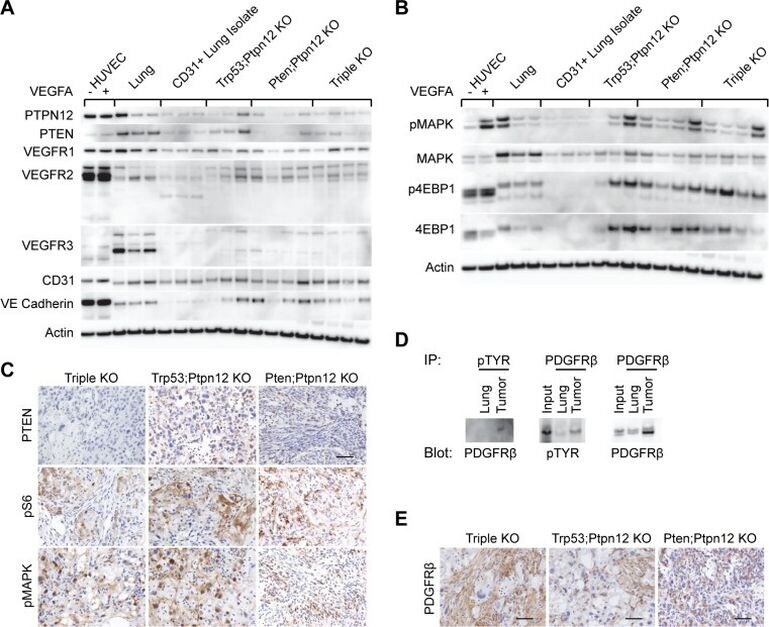
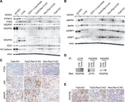
- Figure 2 mTOR and MAPK pathways are activated in murine angiosarcoma ( A ) Western blots analyzing different endothelial cell markers in protein lysates prepared from HUVECs untreated (-) or treated (+) with VEGFA, total lung, endothelial cells isolated from lung using CD31 beads, tumors from both Trp53; Ptpn12 and Pten; Ptpn12 DKO mice, and tumors from TKO mice. ( B ) Western blots using the same protein lysates as in (A) but investigating the indicated signaling molecules. ( C ) Immunohistochemical staining on tissue sections from a representative tumor from each of the indicated mouse genotypes. Scale bar in the top right panel is 50 um and applies to all pictures. ( D ) Anti-phosphotyrosine immunoprecipitations (IP) from control lung and TKO angiosarcoma protein lysates were then probed for PDGFR-beta (left panel). The reciprocal immunoprecipitation and blotting experiment (middle panel) and total PDGFR-beta comparisons (right panel) are shown. The input lane represents 20 ug of protein lysate from the tumor while IPs were performed from 500 ug protein lysate. ( E ) Tumors from the indicated mouse genotypes were stained by IHC for PDGFR-beta. Scale bars are 50 um.