Explore
 Validate
 Learn
Learn Western blot
Antibody data
- Antibody Data
- Antigen structure
- References [0]
- Comments [0]
- Validations
- Western blot [2]
- Immunocytochemistry [1]
Submit
Validation data
Reference
Comment
Report error
- Product number
- PA5-27277 - Provider product page

- Provider
- Invitrogen Antibodies
- Product name
- VEGF Receptor 3 Polyclonal Antibody
- Antibody type
- Polyclonal
- Antigen
- Recombinant protein fragment
- Description
- The recommended shelf life for this product is 1 year from date of receipt.
- Reactivity
- Human, Mouse
- Host
- Rabbit
- Isotype
- IgG
- Vial size
- 100 µL
- Concentration
- 1 mg/mL
- Storage
- Store at 4°C short term. For long term storage, store at -20°C, avoiding freeze/thaw cycles.
No comments: Submit comment
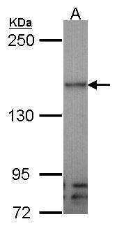
Supportive validation
- Submitted by
- Invitrogen Antibodies (provider)
- Main image

- Experimental details
- Western Blot using VEGF Receptor 3 Polyclonal Antibody (Product # PA5-27277). Sample (30 µg of whole cell lysate). Lane A: Molt-4 .5% SDS PAGE. VEGF Receptor 3 Polyclonal Antibody (Product # PA5-27277) diluted at 1:1,000.
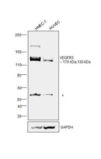
- Submitted by
- Invitrogen Antibodies (provider)
- Main image
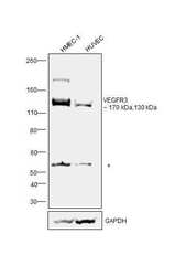
- Experimental details
- Western blot was performed using Anti-VEGF Receptor 3 Polyclonal Antibody (Product # PA5-27277) and a 170 kDa and 130 kDa band corresponding to Vascular endothelial growth factor receptor 3 was observed across cell lines tested. Membrane enriched extracts (40 µg lysate) of HMEC-1 (Lane 1) and HUVEC (Lane 2) were electrophoresed using NuPAGE™ 3-8% Tris-Acetate Protein Gel (Product # EA0378BOX). Resolved proteins were equilibrated with 20% ethanol and then transferred onto a nitrocellulose membrane (Product # IB23002) by iBlot® 2 Dry Blotting System (Product # IB21001). The blot was probed with the primary antibody (1:1000 dilution) and detected by chemiluminescence with Goat anti-Rabbit IgG (H+L) Superclonal™ Recombinant Secondary Antibody, HRP (Product # A27036,1:20000 dilution) using the iBright FL 1000 (Product # A32752). Relative expression of VEGF Receptar 3 was observed to be high in HMEC-1 in comparison to low expression in HUVEC. Chemiluminescent detection was performed using SuperSignal™ West Pico PLUS Chemiluminescent Substrate (Product # 34580).(Ref :DOI: 10.1200/JCO.2004.00.3467).
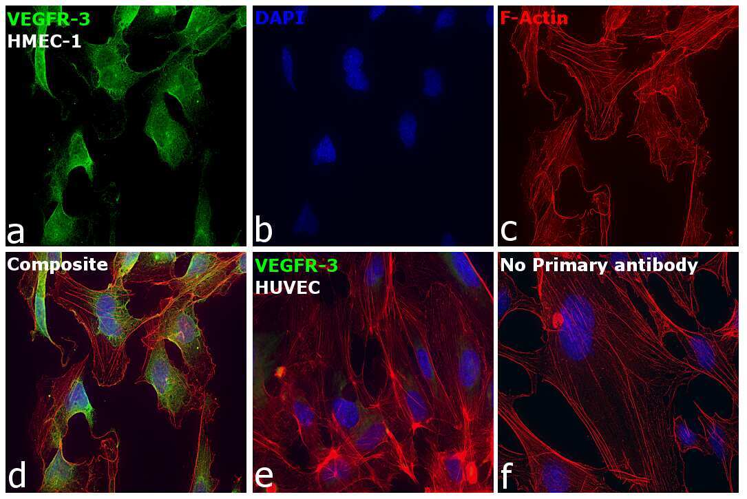
Supportive validation
- Submitted by
- Invitrogen Antibodies (provider)
- Main image
- Experimental details
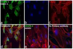
- Immunofluorescence analysis of Vascular endothelial growth factor receptor 3 was performed using 70% confluent log phase HMEC-1 cells. The cells were fixed with 4% paraformaldehyde for 10 minutes, permeabilized with 0.1% Triton™ X-100 for 15 minutes, and blocked with 2% BSA for 45 minutes at room temperature. The cells were labeled with VEGF Receptor 3 Polyclonal Antibody (Product # PA5-27277) at 1:100 dilution in 0.1% BSA, incubated at 4 degree Celsius overnight and then labeled with Donkey anti-Rabbit IgG (H+L) Highly Cross-Adsorbed Secondary Antibody, Alexa Fluor Plus 488 (Product # A32790), (1:2000 dilution), for 45 minutes at room temperature (Panel a: Green). Nuclei (Panel b:Blue) were stained with ProLong™ Diamond Antifade Mountant with DAPI (Product # P36962). F-actin (Panel c: Red) was stained with Rhodamine Phalloidin (Product # R415, 1:300 dilution). Panel d represents the merged image showing Cytoplasmic localization. Panel e represents HUVEC cells having low expression of VEGF Receptar 3. Panel f represents control cells with no primary antibody to assess background. Relative expression of VEGF Receptar 3 was observed to be high in HMEC-1 in comparison to low expression in HUVEC .The images were captured at 60X magnification in EVOS™ M7000 Imaging System (Product # AMF7000) and externally deconvoluted (D.Sage et al. / Methods 115 (2017) 28-41).(Ref :(Ref :DOI: 10.1200/JCO.2004.00.3467).