Antibody data
- Antibody Data
- Antigen structure
- References [1]
- Comments [0]
- Validations
- Immunocytochemistry [3]
- Immunohistochemistry [5]
- Flow cytometry [2]
- Other assay [1]
Submit
Validation data
Reference
Comment
Report error
- Product number
- MA5-34953 - Provider product page

- Provider
- Invitrogen Antibodies
- Product name
- USP21 Monoclonal Antibody (16E1)
- Antibody type
- Monoclonal
- Antigen
- Recombinant full-length protein
- Description
- Positive Control: SH-SY5Y cell lysates, K562 cell lysates, 293T, rat seminal vesicle tissue, human liver tissue, human kidney tissue, human small intestine tissue, mouse heart tissue.
- Reactivity
- Human
- Host
- Mouse
- Isotype
- IgG
- Antibody clone number
- 16E1
- Vial size
- 100 μL
- Concentration
- 2 mg/mL
- Storage
- -20°C, Avoid Freeze/Thaw Cycles, store in dark
Submitted references USP21 promotes self-renewal and tumorigenicity of mesenchymal glioblastoma stem cells by deubiquitinating and stabilizing FOXD1.
Zhang Q, Chen Z, Tang Q, Wang Z, Lu J, You Y, Wang H
Cell death & disease 2022 Aug 16;13(8):712
Cell death & disease 2022 Aug 16;13(8):712
No comments: Submit comment
Supportive validation
- Submitted by
- Invitrogen Antibodies (provider)
- Main image
- Experimental details
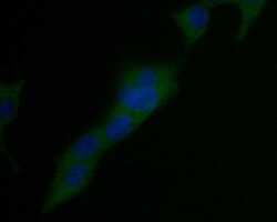
- Immunofluorescent analysis of USP21 in 293T cells (green). Samples were formalin fixed, permeabilized with 0.1% Triton X-100 in TBS (1 hour, room temperature) and blocked with 1% BSA (15 min, room temperature), incubated with USP21 monoclonal antibody (Product # MA5-34953) at a dilution of 1:50 (1 hour, room temperature), and followed by Alexa Fluor 488 Goat anti-Rabbit IgG and DAPI (blue) with a dilution of 1:1000.
- Submitted by
- Invitrogen Antibodies (provider)
- Main image
- Experimental details
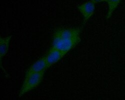
- Immunofluorescent analysis of USP21 in 293T cells (green). Samples were formalin fixed, permeabilized with 0.1% Triton X-100 in TBS (1 hour, room temperature) and blocked with 1% BSA (15 min, room temperature), incubated with USP21 monoclonal antibody (Product # MA5-34953) at a dilution of 1:50 (1 hour, room temperature), and followed by Alexa Fluor 488 Goat anti-Rabbit IgG and DAPI (blue) with a dilution of 1:1000.
- Submitted by
- Invitrogen Antibodies (provider)
- Main image
- Experimental details
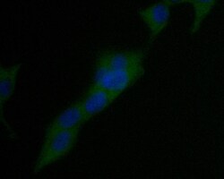
- Immunofluorescent analysis of USP21 in 293T cells (green). Samples were formalin fixed, permeabilized with 0.1% Triton X-100 in TBS (1 hour, room temperature) and blocked with 1% BSA (15 min, room temperature), incubated with USP21 monoclonal antibody (Product # MA5-34953) at a dilution of 1:50 (1 hour, room temperature), and followed by Alexa Fluor 488 Goat anti-Rabbit IgG and DAPI (blue) with a dilution of 1:1000.
Supportive validation
- Submitted by
- Invitrogen Antibodies (provider)
- Main image
- Experimental details
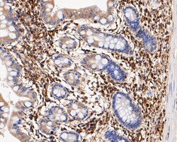
- Immunohistochemistry analysis of USP21 in paraffin-embedded human small intestine tissue. Samples were heat mediated antigen retrieval with Tris-EDTA buffer (pH 8.0-8.4, 20 minutes) and blocked in 5% BSA (30 min, room temperature), incubated with USP21 monoclonal antibody (Product # MA5-34953) at a dilution of 1:200 (30 min, room temperature), and followed by HRP conjugate, DAB and hematoxylin (mounted with DPX).
- Submitted by
- Invitrogen Antibodies (provider)
- Main image
- Experimental details
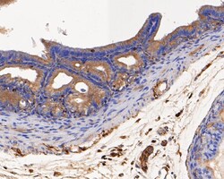
- Immunohistochemistry analysis of USP21 in paraffin-embedded rat seminal vesicle tissue. Samples were heat mediated antigen retrieval with Tris-EDTA buffer (pH 8.0-8.4, 20 minutes) and blocked in 5% BSA (30 min, room temperature), incubated with USP21 monoclonal antibody (Product # MA5-34953) at a dilution of 1:50 (30 min, room temperature), and followed by HRP conjugate, DAB and hematoxylin (mounted with DPX).
- Submitted by
- Invitrogen Antibodies (provider)
- Main image
- Experimental details
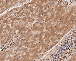
- Immunohistochemistry analysis of USP21 in paraffin-embedded human liver tissue. Samples were heat mediated antigen retrieval with Tris-EDTA buffer (pH 8.0-8.4, 20 minutes) and blocked in 5% BSA (30 min, room temperature), incubated with USP21 monoclonal antibody (Product # MA5-34953) at a dilution of 1:50 (30 min, room temperature), and followed by HRP conjugate, DAB and hematoxylin (mounted with DPX).
- Submitted by
- Invitrogen Antibodies (provider)
- Main image
- Experimental details
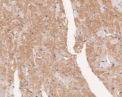
- Immunohistochemistry analysis of USP21 in paraffin-embedded mouse heart tissue. Samples were heat mediated antigen retrieval with Tris-EDTA buffer (pH 8.0-8.4, 20 minutes) and blocked in 5% BSA (30 min, room temperature), incubated with USP21 monoclonal antibody (Product # MA5-34953) at a dilution of 1:50 (30 min, room temperature), and followed by HRP conjugate, DAB and hematoxylin (mounted with DPX).
- Submitted by
- Invitrogen Antibodies (provider)
- Main image
- Experimental details
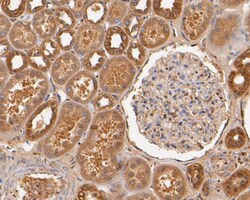
- Immunohistochemistry analysis of USP21 in paraffin-embedded human kidney tissue. Samples were heat mediated antigen retrieval with Tris-EDTA buffer (pH 8.0-8.4, 20 minutes) and blocked in 5% BSA (30 min, room temperature), incubated with USP21 monoclonal antibody (Product # MA5-34953) at a dilution of 1:50 (30 min, room temperature), and followed by HRP conjugate, DAB and hematoxylin (mounted with DPX).
Supportive validation
- Submitted by
- Invitrogen Antibodies (provider)
- Main image
- Experimental details
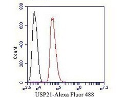
- Flow cytometry of USP21 in SH-SY5Y cells, unlabelled sample (control; cells without incubation with primary antibody; black). Samples were incubated with USP21 monoclonal antibody (Product # MA5-34953) at a dilution of 1:50, followed by Alexa Fluor 488-conjugated Goat anti-Mouse IgG with a dilution of 1:1000 (30 min).
- Submitted by
- Invitrogen Antibodies (provider)
- Main image
- Experimental details
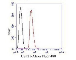
- Flow cytometry of USP21 in SH-SY5Y cells, unlabelled sample (control; cells without incubation with primary antibody; black). Samples were incubated with USP21 monoclonal antibody (Product # MA5-34953) at a dilution of 1:50, followed by Alexa Fluor 488-conjugated Goat anti-Mouse IgG with a dilution of 1:1000 (30 min).
Supportive validation
- Submitted by
- Invitrogen Antibodies (provider)
- Main image
- Experimental details
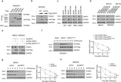
- Fig. 1 USP21 maintains FOXD1 stability. a Co-IP showing that FOXD1 was physically conjugated with USP21 among the predicated proteins(COPS5, SENP3, USP22, USP21). Anti-Flag antibody was used to bind Flag-tagged USP21 WT or C221A. Anti-Myc antibody was used to bind Myc-tagged FOXD1. b Western blotting showing that transiently transfecting of USP21-overexpressing plasmid altered the expression of FOXD1 in a dose-dependent manner. c Western blotting showing that knockdown of USP21 attenuated the protein expression of FOXD1 in U87, U251, T98G and LN229 GBM cell lines. d Western blotting shows that knockdown of USP21 attenuated the expression of FOXD1 in MES 21 and 505 GSCs, whereas treatment with MG-132 (20 muM) abolished the effect of the knockdown of USP21 in MES 21 and 505 GSCs thus increasing the expression of FOXD1. e Western blotting showing that the overexpression of an shRNA-resistant WT, but not C221A mutant, USP21 altered the effect of the knockdown of USP21 in MES 21 and 505 GSCs thus increasing the expression of FOXD1. f Western blotting showing that the overexpression of wild-type USP21 (USP21 WT) but not the catalytically inactive USP21 mutant (USP21 C221A) stabilized FOXD1. *** P < 0.001. g , h Western blotting showing that the knockdown of USP21 in MES 21 ( g ) and 505 ( h ) GSCs resulted in accelerated degradation of FOXD1. *** P < 0.001.
 Explore
 Validate
 Learn
Learn Western blot
 Immunocytochemistry