Explore
 Validate
 Learn
LearnMA5-11950
antibody from Invitrogen Antibodies
Targeting: BCL2L1
Bcl-X, bcl-xL, bcl-xS, BCL2L, BCLX, PPP1R52
 Immunocytochemistry
Antibody data
- Antibody Data
- Antigen structure
- References [14]
- Comments [0]
- Validations
- Immunocytochemistry [9]
- Immunoprecipitation [1]
- Immunohistochemistry [2]
- Flow cytometry [6]
Submit
Validation data
Reference
Comment
Report error
- Product number
- MA5-11950 - Provider product page

- Provider
- Invitrogen Antibodies
- Product name
- Bcl-xL Monoclonal Antibody (7D9)
- Antibody type
- Monoclonal
- Antigen
- Synthetic peptide
- Description
- MA5-11950 targets BCL-XL in FACS, IF, IHC (P) and IP applications. MA5-11950 can be used to stain cells by IF following fixation/permeabilization in either methanol or 4% paraformaldehyde/0.5% octyl-beta-glucoside. The antibody can be used at a lower concentration when fixed and permeabilized by the latter method.
- Reactivity
- Human, Mouse
- Host
- Mouse
- Isotype
- IgG
- Antibody clone number
- 7D9
- Vial size
- 500 μL
- Concentration
- 0.2 mg/mL
- Storage
- 4°C
Submitted references Parthenolide inhibits the proliferation and induces the apoptosis of human uveal melanoma cells.
The anti-cancer effects of Tualang honey in modulating breast carcinogenesis: an experimental animal study.
New diagnostic markers in salivary gland tumors.
Biomarkers in advanced larynx cancer.
Identification of novel target proteins in sebaceous gland carcinoma.
Head and neck squamous cell carcinoma in pregnant women.
Prognostic and predictive markers in medullary thyroid carcinoma.
Biomarkers of parathyroid carcinoma.
MyoD regulates apoptosis of myoblasts through microRNA-mediated down-regulation of Pax3.
Radiotherapy in laryngeal carcinoma: can a panel of 13 markers predict response?
Expression of p53 and Bcl-xL as predictive markers for larynx preservation in advanced laryngeal cancer.
Targeting apoptosis to overcome cisplatin resistance: a translational study in head and neck cancer.
A phase I, pharmacokinetic and biologic correlative study of oblimersen sodium (Genasense, G3139) and irinotecan in patients with metastatic colorectal cancer.
Bcl-2 expression predicts radiotherapy failure in laryngeal cancer.
Che ST, Bie L, Li X, Qi H, Yu P, Zuo L
International journal of ophthalmology 2019;12(10):1531-1538
International journal of ophthalmology 2019;12(10):1531-1538
The anti-cancer effects of Tualang honey in modulating breast carcinogenesis: an experimental animal study.
Ahmed S, Othman NH
BMC complementary and alternative medicine 2017 Apr 11;17(1):208
BMC complementary and alternative medicine 2017 Apr 11;17(1):208
New diagnostic markers in salivary gland tumors.
Schneider S, Kloimstein P, Pammer J, Brannath W, Grasl MCh, Erovic BM
European archives of oto-rhino-laryngology : official journal of the European Federation of Oto-Rhino-Laryngological Societies (EUFOS) : affiliated with the German Society for Oto-Rhino-Laryngology - Head and Neck Surgery 2014 Jul;271(7):1999-2007
European archives of oto-rhino-laryngology : official journal of the European Federation of Oto-Rhino-Laryngological Societies (EUFOS) : affiliated with the German Society for Oto-Rhino-Laryngology - Head and Neck Surgery 2014 Jul;271(7):1999-2007
Biomarkers in advanced larynx cancer.
Bradford CR, Kumar B, Bellile E, Lee J, Taylor J, D'Silva N, Cordell K, Kleer C, Kupfer R, Kumar P, Urba S, Worden F, Eisbruch A, Wolf GT, Teknos TN, Prince ME, Chepeha DB, Hogikyan ND, Moyer JS, Carey TE
The Laryngoscope 2014 Jan;124(1):179-87
The Laryngoscope 2014 Jan;124(1):179-87
Identification of novel target proteins in sebaceous gland carcinoma.
Erovic BM, Al Habeeb A, Harris L, Goldstein DP, Kim D, Ghazarian D, Irish JC
Head & neck 2013 May;35(5):642-8
Head & neck 2013 May;35(5):642-8
Head and neck squamous cell carcinoma in pregnant women.
Eliassen AM, Hauff SJ, Tang AL, Thomas DH, McHugh JB, Walline HM, Stoerker J, Maxwell JH, Worden FP, Eisbruch A, Czerwinski MJ, Papagerakis SM, Chepeha DB, Bradford CR, Hanauer DA, Carey TE, Prince ME
Head & neck 2013 Mar;35(3):335-42
Head & neck 2013 Mar;35(3):335-42
Prognostic and predictive markers in medullary thyroid carcinoma.
Erovic BM, Kim D, Cassol C, Goldstein DP, Irish JC, Asa SL, Mete O
Endocrine pathology 2012 Dec;23(4):232-42
Endocrine pathology 2012 Dec;23(4):232-42
Biomarkers of parathyroid carcinoma.
Erovic BM, Harris L, Jamali M, Goldstein DP, Irish JC, Asa SL, Mete O
Endocrine pathology 2012 Dec;23(4):221-31
Endocrine pathology 2012 Dec;23(4):221-31
MyoD regulates apoptosis of myoblasts through microRNA-mediated down-regulation of Pax3.
Hirai H, Verma M, Watanabe S, Tastad C, Asakura Y, Asakura A
The Journal of cell biology 2010 Oct 18;191(2):347-65
The Journal of cell biology 2010 Oct 18;191(2):347-65
Radiotherapy in laryngeal carcinoma: can a panel of 13 markers predict response?
Wildeman MA, Gibcus JH, Hauptmann M, Begg AC, van Velthuysen ML, Hoebers FJ, Mastik MF, Schuuring E, van der Wal JE, van den Brekel MW
The Laryngoscope 2009 Feb;119(2):316-22
The Laryngoscope 2009 Feb;119(2):316-22
Expression of p53 and Bcl-xL as predictive markers for larynx preservation in advanced laryngeal cancer.
Kumar B, Cordell KG, D'Silva N, Prince ME, Adams ME, Fisher SG, Wolf GT, Carey TE, Bradford CR
Archives of otolaryngology--head & neck surgery 2008 Apr;134(4):363-9
Archives of otolaryngology--head & neck surgery 2008 Apr;134(4):363-9
Targeting apoptosis to overcome cisplatin resistance: a translational study in head and neck cancer.
Bauer JA, Kumar B, Cordell KG, Prince ME, Tran HH, Wolf GT, Chepeha DB, Teknos TN, Wang S, Eisbruch A, Tsien CI, Urba SG, Worden FP, Lee J, Griffith KA, Taylor JM, D'Silva N, Wang SJ, Wolter KG, Henson B, Fisher SG, Carey TE, Bradford CR
International journal of radiation oncology, biology, physics 2007;69(2 Suppl):S106-8
International journal of radiation oncology, biology, physics 2007;69(2 Suppl):S106-8
A phase I, pharmacokinetic and biologic correlative study of oblimersen sodium (Genasense, G3139) and irinotecan in patients with metastatic colorectal cancer.
Mita MM, Ochoa L, Rowinsky EK, Kuhn J, Schwartz G, Hammond LA, Patnaik A, Yeh IT, Izbicka E, Berg K, Tolcher AW
Annals of oncology : official journal of the European Society for Medical Oncology 2006 Feb;17(2):313-21
Annals of oncology : official journal of the European Society for Medical Oncology 2006 Feb;17(2):313-21
Bcl-2 expression predicts radiotherapy failure in laryngeal cancer.
Nix P, Cawkwell L, Patmore H, Greenman J, Stafford N
British journal of cancer 2005 Jun 20;92(12):2185-9
British journal of cancer 2005 Jun 20;92(12):2185-9
No comments: Submit comment
Supportive validation
- Submitted by
- Invitrogen Antibodies (provider)
- Main image
- Experimental details
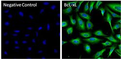
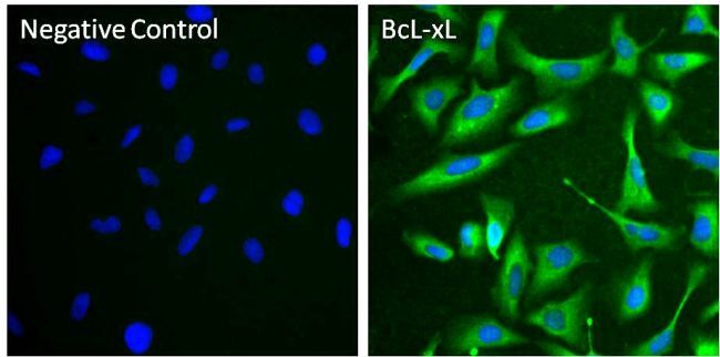
- Immunofluorescent analysis of BcL-xL (green) in U-2 OS cells. Cells were fixed with 4% paraformaldehyde for 15 minutes at room temperature, then permeabilized and blocked with 0.5% Octyl-beta-glucoside (Product # 28310) in 0.3% BSA in PBS for 1 hour at room temperature. Cells were probed with a BcL-xL monoclonal antibody (Product # MA5-11950) at a dilution of 1:50 (right panel), or incubated in blocking buffer as a negative control (left panel) overnight at 4°C. Cells were washed with PBS, and incubated with a DyLight 488 goat anti-mouse IgG secondary antibody (Product # 35502) at a dilution of 1:500 for 1 hour at room temperature. Nuclei (blue) were stained with DAPI (Product # 46190). Images were taken on a Thermo Scientific ToxInsight Instrument at 20X magnification.
- Submitted by
- Invitrogen Antibodies (provider)
- Main image

- Experimental details
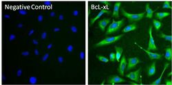
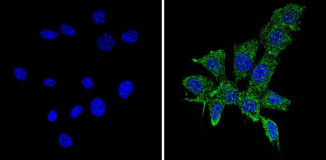
- Immunofluorescent analysis of BcL-xL (green) in U-2 OS cells. Cells were fixed and permeabilized with ice-cold methanol for 10 minutes at room temperature, and blocked with 0.3% BSA in PBS for 1 hour at room temperature. Cells were probed with a BcL-xL monoclonal antibody (Product # MA5-11950) at a dilution of 1:20 (right panel), or incubated in blocking buffer as a negative control (left panel) overnight at 4°C. Cells were washed with PBS, and incubated with a DyLight 488 goat anti-mouse IgG secondary antibody (Product # 35502) at a dilution of 1:500 for 1 hour at room temperature. Nuclei (blue) were stained with DAPI (Product # 46190). Images were taken on a Thermo Scientific ToxInsight Instrument at 20X magnification.
- Submitted by
- Invitrogen Antibodies (provider)
- Main image
- Experimental details
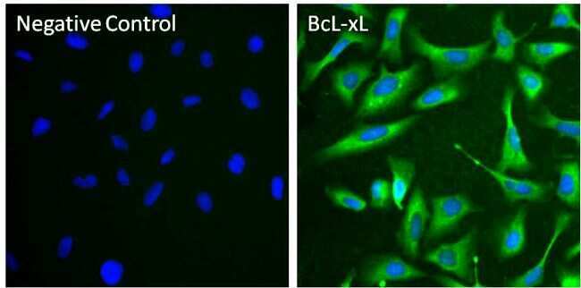
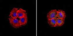
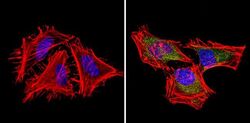
- Immunofluorescent analysis of BCL-XL (green) showing staining in the in the cytoplasm of A431 cells (right) compared to a negative control without primary antibody (left). Formalin-fixed cells were permeabilized with 0.1% Triton X-100 in TBS for 5-10 minutes and blocked with 3% BSA-PBS for 30 minutes at room temperature. Cells were probed with a BCL-XL monoclonal antibody (Product # MA5-11950) in 3% BSA-PBS at a dilution of 1:100 and incubated overnight at 4ºC in a humidified chamber. Cells were washed with PBST and incubated with a DyLight-conjugated secondary antibody in PBS at room temperature in the dark. F-actin (red) was stained with a fluorescent red phalloidin and nuclei (blue) were stained with Hoechst or DAPI. Images were taken at a magnification of 60x.

- Submitted by
- Invitrogen Antibodies (provider)
- Main image

- Experimental details
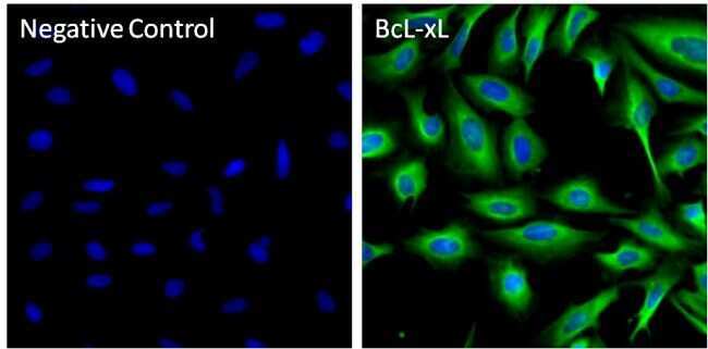
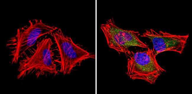
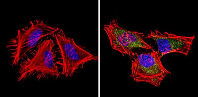
- Immunofluorescent analysis of BCL-XL (green) showing staining in the in the cytoplasm of Hela cells (right) compared to a negative control without primary antibody (left). Formalin-fixed cells were permeabilized with 0.1% Triton X-100 in TBS for 5-10 minutes and blocked with 3% BSA-PBS for 30 minutes at room temperature. Cells were probed with a BCL-XL monoclonal antibody (Product # MA5-11950) in 3% BSA-PBS at a dilution of 1:100 and incubated overnight at 4ºC in a humidified chamber. Cells were washed with PBST and incubated with a DyLight-conjugated secondary antibody in PBS at room temperature in the dark. F-actin (red) was stained with a fluorescent red phalloidin and nuclei (blue) were stained with Hoechst or DAPI. Images were taken at a magnification of 60x.
- Submitted by
- Invitrogen Antibodies (provider)
- Main image

- Experimental details
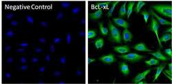
- Immunofluorescent analysis of BCL-XL (green) showing staining in the in the cytoplasm of NIH-3T3 cells (right) compared to a negative control without primary antibody (left). Formalin-fixed cells were permeabilized with 0.1% Triton X-100 in TBS for 5-10 minutes and blocked with 3% BSA-PBS for 30 minutes at room temperature. Cells were probed with a BCL-XL monoclonal antibody (Product # MA5-11950) in 3% BSA-PBS at a dilution of 1:50 and incubated overnight at 4ºC in a humidified chamber. Cells were washed with PBST and incubated with a DyLight-conjugated secondary antibody in PBS at room temperature in the dark. F-actin (red) was stained with a fluorescent red phalloidin and nuclei (blue) were stained with Hoechst or DAPI. Images were taken at a magnification of 60x.

- Submitted by
- Invitrogen Antibodies (provider)
- Main image
- Experimental details
- Immunofluorescent analysis of BcL-xL (green) in U-2 OS cells. Cells were fixed with 4% paraformaldehyde for 15 minutes at room temperature, then permeabilized and blocked with 0.5% Octyl-beta-glucoside (Product # 28310) in 0.3% BSA in PBS for 1 hour at room temperature. Cells were probed with a BcL-xL monoclonal antibody (Product # MA5-11950) at a dilution of 1:50 (right panel), or incubated in blocking buffer as a negative control (left panel) overnight at 4°C. Cells were washed with PBS, and incubated with a DyLight 488 goat anti-mouse IgG secondary antibody (Product # 35502) at a dilution of 1:500 for 1 hour at room temperature. Nuclei (blue) were stained with DAPI (Product # 46190). Images were taken on a Thermo Scientific ToxInsight Instrument at 20X magnification.

- Submitted by
- Invitrogen Antibodies (provider)
- Main image
- Experimental details
- Immunofluorescent analysis of BcL-xL (green) in U-2 OS cells. Cells were fixed and permeabilized with ice-cold methanol for 10 minutes at room temperature, and blocked with 0.3% BSA in PBS for 1 hour at room temperature. Cells were probed with a BcL-xL monoclonal antibody (Product # MA5-11950) at a dilution of 1:20 (right panel), or incubated in blocking buffer as a negative control (left panel) overnight at 4°C. Cells were washed with PBS, and incubated with a DyLight 488 goat anti-mouse IgG secondary antibody (Product # 35502) at a dilution of 1:500 for 1 hour at room temperature. Nuclei (blue) were stained with DAPI (Product # 46190). Images were taken on a Thermo Scientific ToxInsight Instrument at 20X magnification.
- Submitted by
- Invitrogen Antibodies (provider)
- Main image
- Experimental details
- Immunofluorescent analysis of BCL-XL (green) showing staining in the in the cytoplasm of Hela cells (right) compared to a negative control without primary antibody (left). Formalin-fixed cells were permeabilized with 0.1% Triton X-100 in TBS for 5-10 minutes and blocked with 3% BSA-PBS for 30 minutes at room temperature. Cells were probed with a BCL-XL monoclonal antibody (Product # MA5-11950) in 3% BSA-PBS at a dilution of 1:100 and incubated overnight at 4ºC in a humidified chamber. Cells were washed with PBST and incubated with a DyLight-conjugated secondary antibody in PBS at room temperature in the dark. F-actin (red) was stained with a fluorescent red phalloidin and nuclei (blue) were stained with Hoechst or DAPI. Images were taken at a magnification of 60x.
- Submitted by
- Invitrogen Antibodies (provider)
- Main image

- Experimental details
- Immunofluorescent analysis of BCL-XL (green) showing staining in the in the cytoplasm of NIH-3T3 cells (right) compared to a negative control without primary antibody (left). Formalin-fixed cells were permeabilized with 0.1% Triton X-100 in TBS for 5-10 minutes and blocked with 3% BSA-PBS for 30 minutes at room temperature. Cells were probed with a BCL-XL monoclonal antibody (Product # MA5-11950) in 3% BSA-PBS at a dilution of 1:50 and incubated overnight at 4ºC in a humidified chamber. Cells were washed with PBST and incubated with a DyLight-conjugated secondary antibody in PBS at room temperature in the dark. F-actin (red) was stained with a fluorescent red phalloidin and nuclei (blue) were stained with Hoechst or DAPI. Images were taken at a magnification of 60x.
Supportive validation
- Submitted by
- Invitrogen Antibodies (provider)
- Main image
- Experimental details
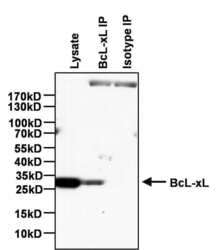
- Immunoprecipitation of BcL-xL was performed on U-2 OS cells. Antigen-antibody complexes were formed by incubating 750 µg of U-2 OS whole cell lysate with 4 µg of a BcL-xL monoclonal antibody (Product # MA5-11950) or Mouse IgG2a isotype control (Product # MA1-10418) overnight on a rocking platform at 4°C. The immune complexes were captured on 100 µL Protein A/G Agarose (Product # 20421), washed extensively, and eluted with 5X Lane Marker Reducing Sample Buffer (Product # 39000). The IP samples and 25 µg of U-2 OS whole cell lysate (as a positive control for Western blot detection) were resolved on a 4-20% Tris-HCl polyacrylamide gel, transferred to a PVDF membrane, and blocked with 5% BSA/TBS-0.1%Tween for at least 1 hour. The membrane was probed with a BcL-xL rabbit monoclonal antibody (Product # MA5-15142) at a dilution of 1:1000 overnight rotating at 4°C, washed in TBST, and probed with an HRP-conjugated mouse anti-rabbit light chain secondary antibody at a dilution of 1:40,000 for at least 1 hour. Chemiluminescent detection was performed using SuperSignal West Pico (Product # 34080).

Supportive validation
- Submitted by
- Invitrogen Antibodies (provider)
- Main image
- Experimental details
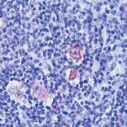
- Formalin-fixed, paraffin-embedded human Hodgkins lymphoma stained with Bcl-X antibody using peroxidase-conjugate and AEC chromogen. Note cytoplasmic staining of Hodgkins cells.

- Submitted by
- Invitrogen Antibodies (provider)
- Main image
- Experimental details
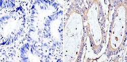
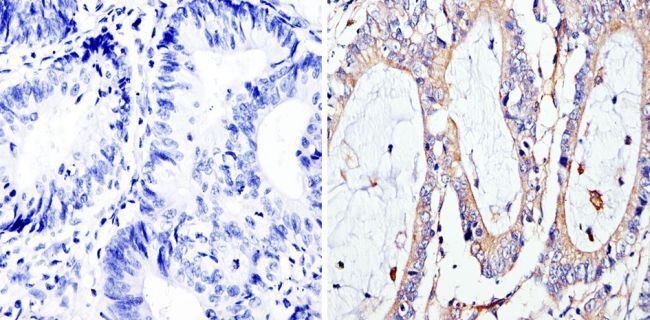
- Immunohistochemistry analysis of BCL-XL showing staining in the cytoplasm of paraffin-embedded human colon carcinoma (right) compared to a negative control without primary antibody (left). To expose target proteins, antigen retrieval was performed using 10mM sodium citrate (pH 6.0), microwaved for 8-15 min. Following antigen retrieval, tissues were blocked in 3% H2O2-methanol for 15 min at room temperature, washed with ddH2O and PBS, and then probed with a BCL-XL monoclonal antibody (Product # MA5-11950) diluted in 3% BSA-PBS at a dilution of 1:100 overnight at 4°C in a humidified chamber. Tissues were washed extensively in PBST and detection was performed using an HRP-conjugated secondary antibody followed by colorimetric detection using a DAB kit. Tissues were counterstained with hematoxylin and dehydrated with ethanol and xylene to prep for mounting.
Supportive validation
- Submitted by
- Invitrogen Antibodies (provider)
- Main image
- Experimental details
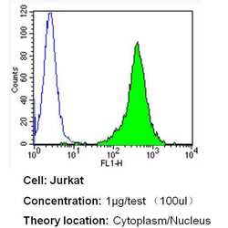
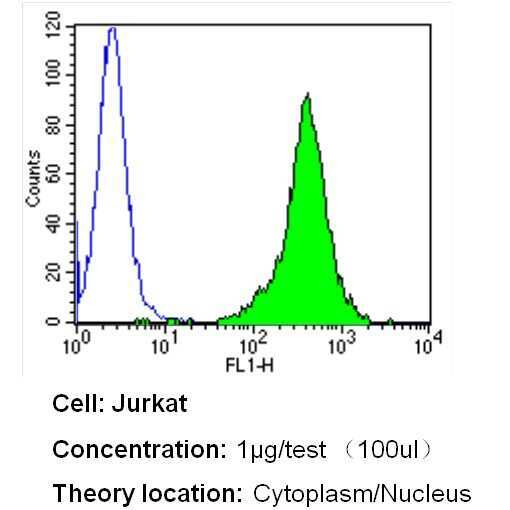
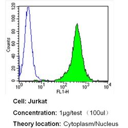
- Flow cytometry analysis of BCL-XL in Jurkat cells (green) compared to an isotype control (blue). Cells were harvested, adjusted to a concentration of 1-5x10^6 cells/mL, fixed with 2% paraformaldehyde and washed with PBS. Cells were blocked with a 2% solution of BSA-PBS for 30 min at room temperature and incubated with a BCL-XL monoclonal antibody (Product # MA5-11950) at a dilution of 1 µg/test for 40 min at room temperature. Cells were then incubated for 40 min at room temperature in the dark using a Dylight 488-conjugated secondary antibody and re-suspended in PBS for FACS analysis.
- Submitted by
- Invitrogen Antibodies (provider)
- Main image
- Experimental details
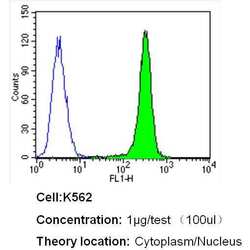
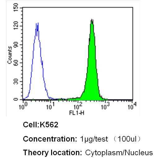
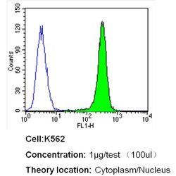
- Flow cytometry analysis of BCL-XL in K562 cells (green) compared to an isotype control (blue). Cells were harvested, adjusted to a concentration of 1-5x10^6 cells/mL, fixed with 2% paraformaldehyde and washed with PBS. Cells were blocked with a 2% solution of BSA-PBS for 30 min at room temperature and incubated with a BCL-XL monoclonal antibody (Product # MA5-11950) at a dilution of 1 µg/test for 40 min at room temperature. Cells were then incubated for 40 min at room temperature in the dark using a Dylight 488-conjugated secondary antibody and re-suspended in PBS for FACS analysis.
- Submitted by
- Invitrogen Antibodies (provider)
- Main image

- Experimental details
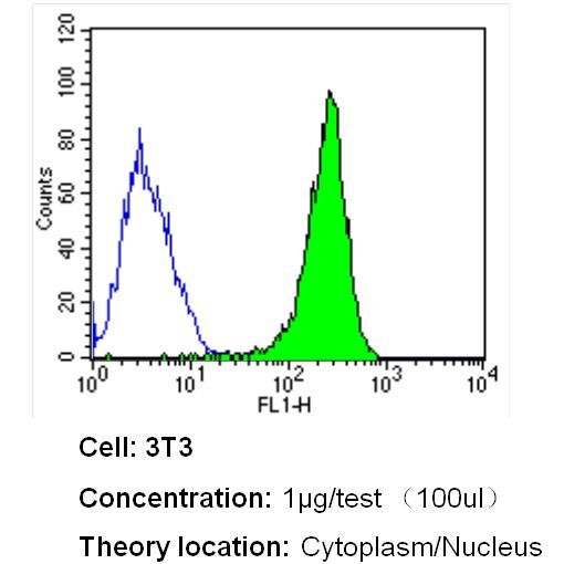
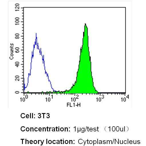
- Flow cytometry analysis of BCL-XL in NIH-3T3 cells (green) compared to an isotype control (blue). Cells were harvested, adjusted to a concentration of 1-5x10^6 cells/mL, fixed with 2% paraformaldehyde and washed with PBS. Cells were blocked with a 2% solution of BSA-PBS for 30 min at room temperature and incubated with a BCL-XL monoclonal antibody (Product # MA5-11950) at a dilution of 1 µg/test for 40 min at room temperature. Cells were then incubated for 40 min at room temperature in the dark using a Dylight 488-conjugated secondary antibody and re-suspended in PBS for FACS analysis.
- Submitted by
- Invitrogen Antibodies (provider)
- Main image
- Experimental details
- Flow cytometry analysis of BCL-XL in Jurkat cells (green) compared to an isotype control (blue). Cells were harvested, adjusted to a concentration of 1-5x10^6 cells/mL, fixed with 2% paraformaldehyde and washed with PBS. Cells were blocked with a 2% solution of BSA-PBS for 30 min at room temperature and incubated with a BCL-XL monoclonal antibody (Product # MA5-11950) at a dilution of 1 µg/test for 40 min at room temperature. Cells were then incubated for 40 min at room temperature in the dark using a Dylight 488-conjugated secondary antibody and re-suspended in PBS for FACS analysis.

- Submitted by
- Invitrogen Antibodies (provider)
- Main image
- Experimental details
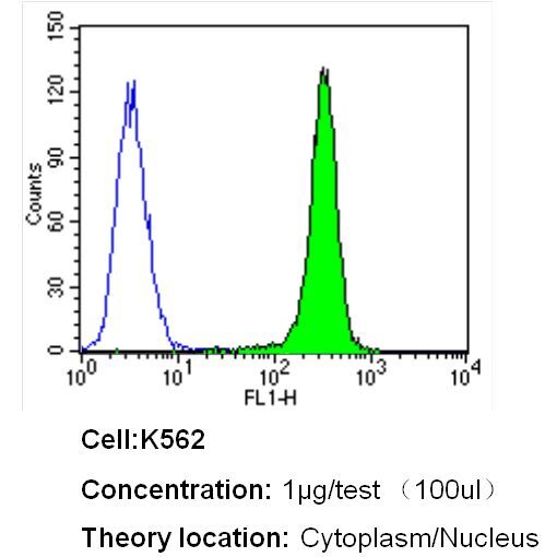
- Flow cytometry analysis of BCL-XL in K562 cells (green) compared to an isotype control (blue). Cells were harvested, adjusted to a concentration of 1-5x10^6 cells/mL, fixed with 2% paraformaldehyde and washed with PBS. Cells were blocked with a 2% solution of BSA-PBS for 30 min at room temperature and incubated with a BCL-XL monoclonal antibody (Product # MA5-11950) at a dilution of 1 µg/test for 40 min at room temperature. Cells were then incubated for 40 min at room temperature in the dark using a Dylight 488-conjugated secondary antibody and re-suspended in PBS for FACS analysis.
- Submitted by
- Invitrogen Antibodies (provider)
- Main image

- Experimental details
- Flow cytometry analysis of BCL-XL in NIH-3T3 cells (green) compared to an isotype control (blue). Cells were harvested, adjusted to a concentration of 1-5x10^6 cells/mL, fixed with 2% paraformaldehyde and washed with PBS. Cells were blocked with a 2% solution of BSA-PBS for 30 min at room temperature and incubated with a BCL-XL monoclonal antibody (Product # MA5-11950) at a dilution of 1 µg/test for 40 min at room temperature. Cells were then incubated for 40 min at room temperature in the dark using a Dylight 488-conjugated secondary antibody and re-suspended in PBS for FACS analysis.