 Explore
 Validate
 Learn
Learn Western blot
 Immunocytochemistry
Antibody data
- Antibody Data
- Antigen structure
- References [0]
- Comments [0]
- Validations
- Immunocytochemistry [4]
- Immunoprecipitation [1]
- Immunohistochemistry [4]
- Other assay [1]
Submit
Validation data
Reference
Comment
Report error
- Product number
- PA5-84240 - Provider product page

- Provider
- Invitrogen Antibodies
- Product name
- NDUFS2 Polyclonal Antibody
- Antibody type
- Polyclonal
- Antigen
- Recombinant protein fragment
- Description
- Immunogen sequence: KVDDAKVSPP KRAEMKTSME SLIHHFKLYT EGYQVPPGAT YTAIEAPKGE FGVYLVSDGS SRPYRCKIKA PGFAHLAGLD
- Reactivity
- Human
- Host
- Rabbit
- Isotype
- IgG
- Vial size
- 100 μL
- Concentration
- 0.1 mg/mL
- Storage
- Store at 4°C short term. For long term storage, store at -20°C, avoiding freeze/thaw cycles.
No comments: Submit comment
Supportive validation
- Submitted by
- Invitrogen Antibodies (provider)
- Main image
- Experimental details
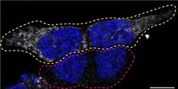
- Immunofluorescence of NDUFS2 was performed using HAP1 wild-type and NDUFS2 KO cells that were transfected with a green or a far red fluorescent dye, respectively. Post-transfection, WT and KO cells were mixed and plated to a 1:1 ratio on coverslips as a mosaic and incubated for 24 hrs. Cells were fixed in 4% PFA (in PBS) for 15 min; cells were permeabilized with 0.1% Triton X-100 for 10 min at RT and blocked with PBS with 5% BSA, 5% goat serum, and 0.01% Triton X-100 for 30 min. Cells were stained with the NDUFS2 polyclonal antibody (Product # PA5-84240) at a 1:100 dilution overnight at 4°C. Secondary antibody incubation was performed using 0.5 µg/mL of Goat anti-Rabbit IgG (H+L) Highly Cross-Adsorbed Secondary Antibody, Alexa Fluor 555 antibody (Product # A21429) together with DAPI. Imaging was performed with a 40X oil objective and analysis was performed using Image J. Cell image represents a single focal plane; WT and KO cells are outlined with a yellow (WT) or magenta (KO) dashed line. Data courtesy of YCharOS Inc., an open science company with the mission of characterizing commercially available antibodies using knockout validation.

- Submitted by
- Invitrogen Antibodies (provider)
- Main image
- Experimental details
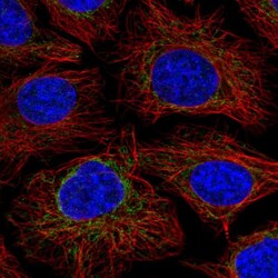
- Immunofluorescent analysis of NDUFS2 in HeLa cells using a NDUFS2 polyclonal antibody (Product # PA5-84240). The analysis shows localization to mitochondria.

- Submitted by
- Invitrogen Antibodies (provider)
- Main image

- Experimental details
- Immunofluorecent analysis of NDUFS2 in human cell line HeLa using NDUFS2 Polyclonal Antibody (Product # PA5-84240). Staining shows localization to mitochondria.

- Submitted by
- Invitrogen Antibodies (provider)
- Main image

- Experimental details
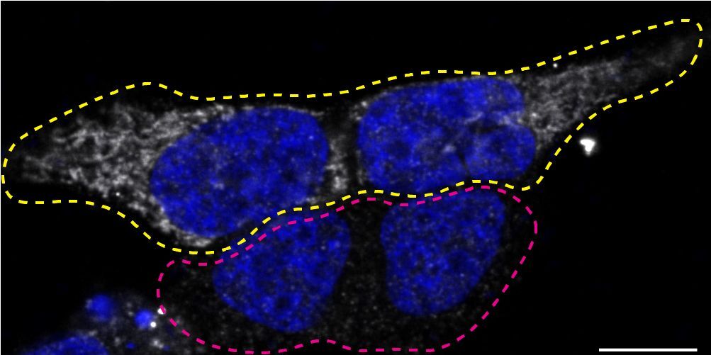
- Immunofluorescence of NDUFS2 was performed using HAP1 wild-type and NDUFS2 KO cells that were transfected with a green or a far red fluorescent dye, respectively. Post-transfection, WT and KO cells were mixed and plated to a 1:1 ratio on coverslips as a mosaic and incubated for 24 hrs. Cells were fixed in 4% PFA (in PBS) for 15 min; cells were permeabilized with 0.1% Triton X-100 for 10 min at RT and blocked with PBS with 5% BSA, 5% goat serum, and 0.01% Triton X-100 for 30 min. Cells were stained with the NDUFS2 polyclonal antibody (Product # PA5-84240) at a 1:100 dilution overnight at 4°C. Secondary antibody incubation was performed using 0.5 µg/mL of Goat anti-Rabbit IgG (H+L) Highly Cross-Adsorbed Secondary Antibody, Alexa Fluor 555 antibody (Product # A21429) together with DAPI. Imaging was performed with a 40X oil objective and analysis was performed using Image J. Cell image represents a single focal plane; WT and KO cells are outlined with a yellow (WT) or magenta (KO) dashed line. Data courtesy of YCharOS Inc., an open science company with the mission of characterizing commercially available antibodies using knockout validation.
Supportive validation
- Submitted by
- Invitrogen Antibodies (provider)
- Main image
- Experimental details
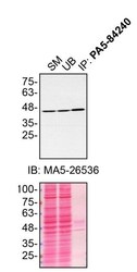
- Immunoprecipitation of NDUFS2 was performed on HAP1 cell lysates. Antibody-bead conjugates were prepared by adding 2 µg of NDUFS2 polyclonal antibody (Product # PA5-84240) with 30 µL of protein A-Sepharose beads and rocked overnight at 4°C. 500 µg of lysate was incubated with an antibody-bead conjugate for 2 hours at 4°C. Following centrifugation and multiple washes, 10% starting material (SM), 10% unbound fraction (UB) and immunoprecipitated fraction (IP) were processed for immunoblot using the same antibody. Ponceau stained transfer of blot is shown. Data courtesy of YCharOS Inc., an open science company with the mission of characterizing commercially available antibodies using knockout validation.

Supportive validation
- Submitted by
- Invitrogen Antibodies (provider)
- Main image
- Experimental details
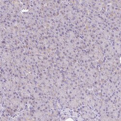
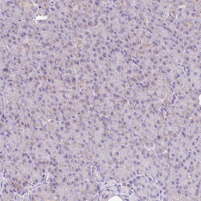
- Immunohistochemical analysis of NDUFS2 in human pancreas using NDUFS2 Polyclonal Antibody (Product # PA5-84240) shows very weak granular cytoplasmic positivity in exocrine glandular cells.
- Submitted by
- Invitrogen Antibodies (provider)
- Main image
- Experimental details
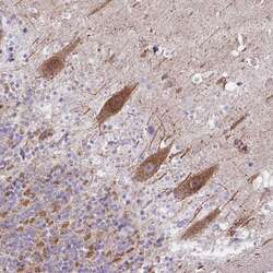
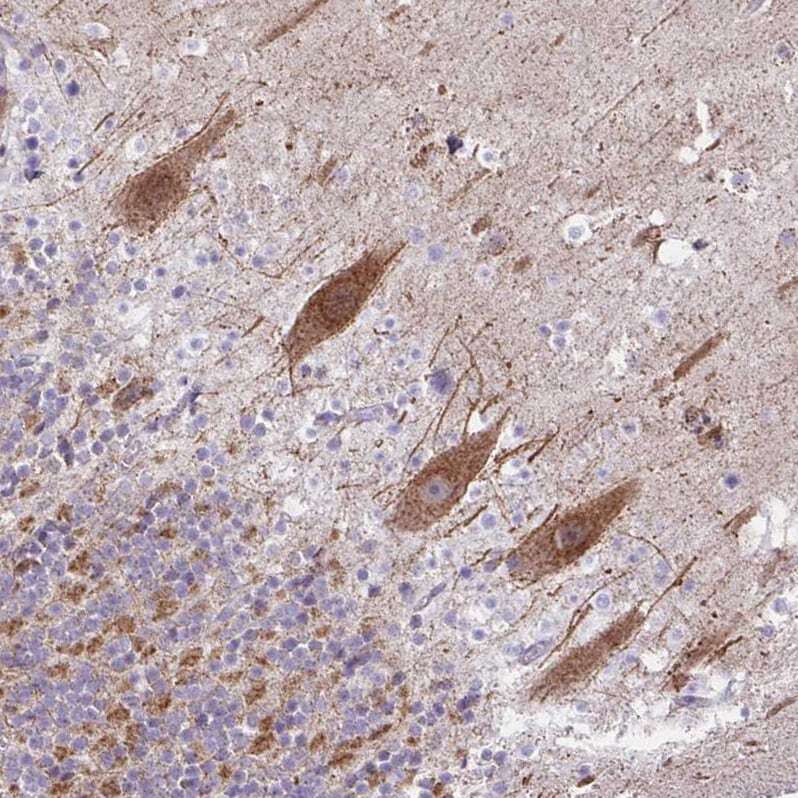
- Immunohistochemical analysis of NDUFS2 in human cerebellum using NDUFS2 Polyclonal Antibody (Product # PA5-84240) shows moderate granular cytoplasmic positivity in Purkinje cells.
- Submitted by
- Invitrogen Antibodies (provider)
- Main image

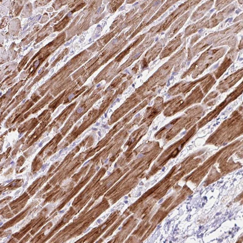
- Experimental details
- Immunohistochemical analysis of NDUFS2 in human heart muscle using NDUFS2 Polyclonal Antibody (Product # PA5-84240) shows strong cytoplasmic positivity in cardiomyocytes.
- Submitted by
- Invitrogen Antibodies (provider)
- Main image
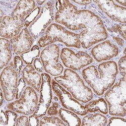
- Experimental details
- Immunohistochemical analysis of NDUFS2 in human kidney using NDUFS2 Polyclonal Antibody (Product # PA5-84240) shows moderate granular cytoplasmic positivity in cells in tubules.

Supportive validation
- Submitted by
- Invitrogen Antibodies (provider)
- Main image

- Experimental details
- Immunoprecipitation of NDUFS2 was performed on HAP1 cell lysates. Antibody-bead conjugates were prepared by adding 2 µg of NDUFS2 polyclonal antibody (Product # PA5-84240) with 30 µL of protein A-Sepharose beads and rocked overnight at 4°C. 500 µg of lysate was incubated with an antibody-bead conjugate for 2 hours at 4°C. Following centrifugation and multiple washes, 10% starting material (SM), 10% unbound fraction (UB) and immunoprecipitated fraction (IP) were processed for immunoblot using the same antibody. Ponceau stained transfer of blot is shown. Data courtesy of YCharOS Inc., an open science company with the mission of characterizing commercially available antibodies using knockout validation.
