 Explore
 Validate
 Learn
Learn Western blot
 Immunoprecipitation
Antibody data
- Antibody Data
- Antigen structure
- References [2]
- Comments [0]
- Validations
- Western blot [3]
- Immunocytochemistry [1]
- Immunohistochemistry [1]
- Other assay [3]
Submit
Validation data
Reference
Comment
Report error
- Product number
- PA5-22364 - Provider product page

- Provider
- Invitrogen Antibodies
- Product name
- NDUFS2 Polyclonal Antibody
- Antibody type
- Polyclonal
- Antigen
- Recombinant full-length protein
- Description
- Recommended positive controls: 293T, A431, Jurkat, Raji, mouse brain, rat brain. Predicted reactivity: Mouse (95%), Rat (94%), Zebrafish (84%), Xenopus laevis (90%), Rhesus Monkey (98%), Chimpanzee (100%), Bovine (94%). Store product as a concentrated solution. Centrifuge briefly prior to opening the vial.
- Reactivity
- Human, Mouse, Rat
- Host
- Rabbit
- Isotype
- IgG
- Vial size
- 100 μL
- Concentration
- 0.43 mg/mL
- Storage
- Store at 4°C short term. For long term storage, store at -20°C, avoiding freeze/thaw cycles.
Submitted references Metabolic rescue ameliorates mitochondrial encephalo-cardiomyopathy in murine and human iPSC models of Leigh syndrome.
NDUFS3 depletion permits complex I maturation and reveals TMEM126A/OPA7 as an assembly factor binding the ND4-module intermediate.
Yoon JY, Daneshgar N, Chu Y, Chen B, Hefti M, Vikram A, Irani K, Song LS, Brenner C, Abel ED, London B, Dai DF
Clinical and translational medicine 2022 Jul;12(7):e954
Clinical and translational medicine 2022 Jul;12(7):e954
NDUFS3 depletion permits complex I maturation and reveals TMEM126A/OPA7 as an assembly factor binding the ND4-module intermediate.
D'Angelo L, Astro E, De Luise M, Kurelac I, Umesh-Ganesh N, Ding S, Fearnley IM, Gasparre G, Zeviani M, Porcelli AM, Fernandez-Vizarra E, Iommarini L
Cell reports 2021 Apr 20;35(3):109002
Cell reports 2021 Apr 20;35(3):109002
No comments: Submit comment
Supportive validation
- Submitted by
- Invitrogen Antibodies (provider)
- Main image
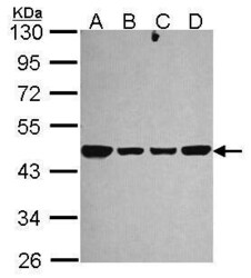
- Experimental details
- Western Blot using NDUFS2 Polyclonal Antibody (Product # PA5-22364). Sample (30 µg of whole cell lysate). Lane A: 293T. Lane B: A431. Lane C: JurKat. Lane D: Raji. 10% SDS PAGE. NDUFS2 Polyclonal Antibody (Product # PA5-22364) diluted at 1:1,000.
- Submitted by
- Invitrogen Antibodies (provider)
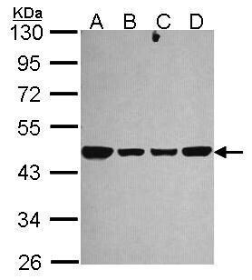
- Main image

- Experimental details
- Western Blot using NDUFS2 Polyclonal Antibody (Product # PA5-22364). Sample (50 µg of whole cell lysate). Lane A: Mouse brain. 10% SDS PAGE. NDUFS2 Polyclonal Antibody (Product # PA5-22364) diluted at 1:10,000.
- Submitted by
- Invitrogen Antibodies (provider)
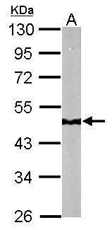
- Main image
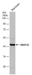
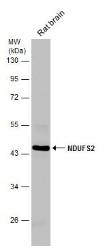
- Experimental details
- Western Blot analysis of NDUFS2 was performed by separating 50 µg of rat tissue extract by 10% SDS-PAGE. Proteins were transferred to a membrane and probed with a NDUFS2 Polyclonal Antibody (Product # PA5-22364) at a dilution of 1:1,000.
Supportive validation
- Submitted by
- Invitrogen Antibodies (provider)
- Main image
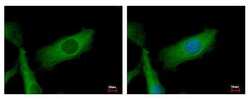
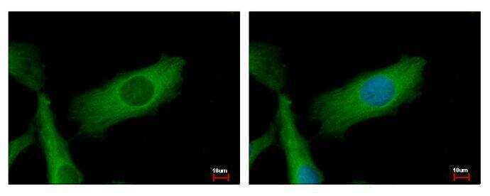
- Experimental details
- NDUFS2 Polyclonal Antibody detects NDUFS2 protein at mitochondria by immunofluorescent analysis. Sample: HeLa cells were fixed in 2% paraformaldehyde/culture medium at 37ºC for 30 min. Green: NDUFS2 protein stained by NDUFS2 Polyclonal Antibody (Product # PA5-22364) diluted at 1:500. Blue: Hoechst 33343 staining.
Supportive validation
- Submitted by
- Invitrogen Antibodies (provider)
- Main image
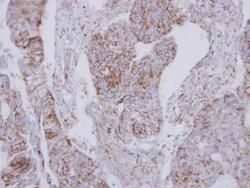
- Experimental details
- Immunohistochemical analysis of paraffin-embedded human colon carcinoma, using NDUFS2 (Product # PA5-22364) antibody at 1:250 dilution. Antigen Retrieval: EDTA based buffer, pH 8.0, 15 min.

Supportive validation
- Submitted by
- Invitrogen Antibodies (provider)
- Main image
- Experimental details
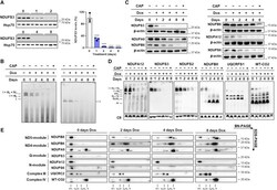
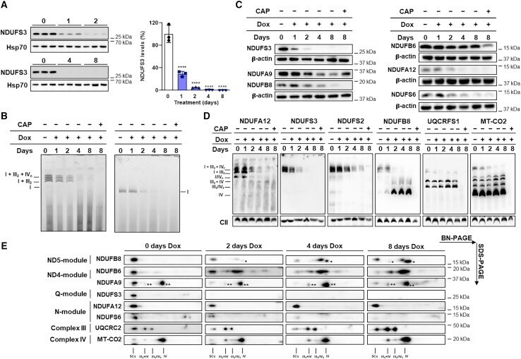
- Figure 2 Persistence of functional respiratory CI during repression of NDUFS3 (A) Immunodetection of NDUFS3 on western blots of whole-cell lysates from 143B -/- NDUFS3 cells treated with 100 ng/mL doxycycline (Dox) for 0 (untreated), 1, 2, 4, and 8 days (n = 3). NDUFS3 band intensities were quantified by densitometry and normalized to the signal of Hsp70, used as loading control. The mean values of the treated cells were referred to those of untreated (control) set to 100%. Data are means +- SD. **** p < 0.0001 treated versus untreated; one-way ANOVA with Sidak''s multiple comparisons test. (B) CI-IGA of enriched mitochondrial fractions from repressed 143B -/-NDUFS3 , solubilized either with digitonin or DDM and separated by BN-PAGE. SCs include CI+III 2 and respirasomes (I+III 2 +IV 1-n ). (C) Immunodetection of CI subunits on western blots of 143B -/-NDUFS3 cells, treated either with 100 ng/mL Dox for 0 (untreated), 1, 2, 4, and 8 days or simultaneously with 100 ng/mL Dox and 50 mug/mL CAP for 8 days, resolved by SDS-PAGE. beta-actin was used as loading control. (D) Immunodetection of CI subunits NDUFA12 (N-module), NDUFS2 (Q-module), NDUFS3 (Q-module), NDUFB8 (ND5-module), CIII subunit UQCRFS1, and CIV subunit MT-CO2, in western blots of mitochondrial fractions from 143B -/-NDUFS3 , treated either with 100 ng/mL Dox for 0 (untreated), 1, 2, 4, and 8 days or simultaneously with 100 ng/mL Dox and 50 mug/mL CAP for 8 days, solubilized with digitonin and separated by BN-PAGE.
- Submitted by
- Invitrogen Antibodies (provider)
- Main image

- Experimental details
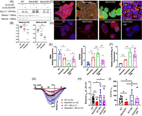
- FIGURE 4 Acetylation of K-1479- Na V 1.5 in mitochondrial complex I-deficient cells decrease sodium current. (A) Western blot for Na V 1.5 protein in 293 cells lacking Ndufs4 (Ndufs4 KO) or Ndufs2 (Ndufs2 KO) which were transfected with plasmid carrying WT or mutant Scn5a gene (to produce WT or acetylation-site mutant Na V 1.5 proteins). (B) pH of conditioned-media from 293, Ndufs4 KO, and Ndufs2 KO cells at 24 or 48 h after plating ( n = 6/group). (C) Live staining of 293 (WT), Ndufs4 KO, and Ndufs2 KO cells with markers of mitochondrial membrane potential (TMRE), total cellular ROS (DCFDA) and Hoechst (blue). (D) Representative fluorescence images of acetylation of Na V 1.5 residue K1479. (E) Quantitation of results in panel C ( n = 5-10/group). (F) Quantitation of results in (D). ( n = 4-12/group). (G) Patch clamp results of I Na for WT and Ndufs2 KO cells with or without NR treatment. Quantitation of (H) relative I Na density at -20 mV and (I) AUC for the indicated groups in panel (G). n = 7-19. Data are mean +- s.e.m. of biologically independent samples. Statistical significance was determined by ANOVA and post-hoc analyses. * p < .05, ** p < .01, ns not significant
- Submitted by
- Invitrogen Antibodies (provider)
- Main image

- Experimental details
- 4 FIGURE Acetylation of K-1479- Na V 1.5 in mitochondrial complex I-deficient cells decrease sodium current. (A) Western blot for Na V 1.5 protein in 293 cells lacking Ndufs4 (Ndufs4 KO) or Ndufs2 (Ndufs2 KO) which were transfected with plasmid carrying WT or mutant Scn5a gene (to produce WT or acetylation-site mutant Na V 1.5 proteins). (B) pH of conditioned-media from 293, Ndufs4 KO, and Ndufs2 KO cells at 24 or 48 h after plating ( n = 6/group). (C) Live staining of 293 (WT), Ndufs4 KO, and Ndufs2 KO cells with markers of mitochondrial membrane potential (TMRE), total cellular ROS (DCFDA) and Hoechst (blue). (D) Representative fluorescence images of acetylation of Na V 1.5 residue K1479. (E) Quantitation of results in panel C ( n = 5-10/group). (F) Quantitation of results in (D). ( n = 4-12/group). (G) Patch clamp results of I Na for WT and Ndufs2 KO cells with or without NR treatment. Quantitation of (H) relative I Na density at -20 mV and (I) AUC for the indicated groups in panel (G). n = 7-19. Data are mean +- s.e.m. of biologically independent samples. Statistical significance was determined by ANOVA and post-hoc analyses. * p < .05, ** p < .01, ns not significant
