Explore
 Validate
 Learn
Learn Western blot
Antibody data
- Antibody Data
- Antigen structure
- References [4]
- Comments [0]
- Validations
- Western blot [4]
- Immunocytochemistry [1]
- Immunoprecipitation [1]
- Immunohistochemistry [1]
Submit
Validation data
Reference
Comment
Report error
- Product number
- GTX128427 - Provider product page

- Provider
- GeneTex
- Product name
- ATG9A antibody
- Antibody type
- Polyclonal
- Reactivity
- Human, Mouse, Rat
- Host
- Rabbit
Submitted references Attenuation of cGAS-STING signaling is mediated by a p62/SQSTM1-dependent autophagy pathway activated by TBK1.
PI3K-C2α knockdown decreases autophagy and maturation of endocytic vesicles.
Bif-1 deficiency impairs lipid homeostasis and causes obesity accompanied by insulin resistance.
Autophagy initiation by ULK complex assembly on ER tubulovesicular regions marked by ATG9 vesicles.
Prabakaran T, Bodda C, Krapp C, Zhang BC, Christensen MH, Sun C, Reinert L, Cai Y, Jensen SB, Skouboe MK, Nyengaard JR, Thompson CB, Lebbink RJ, Sen GC, van Loo G, Nielsen R, Komatsu M, Nejsum LN, Jakobsen MR, Gyrd-Hansen M, Paludan SR
The EMBO journal 2018 Apr 13;37(8)
The EMBO journal 2018 Apr 13;37(8)
PI3K-C2α knockdown decreases autophagy and maturation of endocytic vesicles.
Merrill NM, Schipper JL, Karnes JB, Kauffman AL, Martin KR, MacKeigan JP
PloS one 2017;12(9):e0184909
PloS one 2017;12(9):e0184909
Bif-1 deficiency impairs lipid homeostasis and causes obesity accompanied by insulin resistance.
Liu Y, Takahashi Y, Desai N, Zhang J, Serfass JM, Shi YG, Lynch CJ, Wang HG
Scientific reports 2016 Feb 9;6:20453
Scientific reports 2016 Feb 9;6:20453
Autophagy initiation by ULK complex assembly on ER tubulovesicular regions marked by ATG9 vesicles.
Karanasios E, Walker SA, Okkenhaug H, Manifava M, Hummel E, Zimmermann H, Ahmed Q, Domart MC, Collinson L, Ktistakis NT
Nature communications 2016 Aug 11;7:12420
Nature communications 2016 Aug 11;7:12420
No comments: Submit comment
Supportive validation
- Submitted by
- GeneTex (provider)
- Main image

- Experimental details
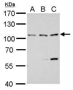
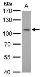
- ATG9A antibody detects ATG9A protein by western blot analysis.A. 30 ?g 293T whole cell lysate/extractB. 30 ?g A431 whole cell lysate/extractC. 30 ?g HeLa whole cell lysate/extract7.5% SDS-PAGEATG9A antibody (GTX128427) dilution: 1:1000 The HRP-conjugated anti-rabbit IgG antibody (GTX213110-01) was used to detect the primary antibody.
- Submitted by
- GeneTex (provider)
- Main image
- Experimental details
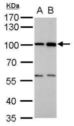
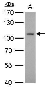
- ATG9A antibody detects ATG9A protein by western blot analysis.A. 30 ?g PC-12 whole cell lysate/extractB. 30 ?g Rat-2 whole cell lysate/extract7.5% SDS-PAGEATG9A antibody (GTX128427) dilution: 1:1000 The HRP-conjugated anti-rabbit IgG antibody (GTX213110-01) was used to detect the primary antibody.
- Submitted by
- GeneTex (provider)
- Main image
- Experimental details
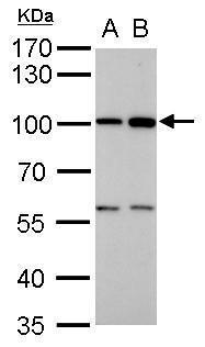
- ATG9A antibody detects ATG9A protein by western blot analysis.A. 30 ?g BCL-1 whole cell lysate/extract7.5% SDS-PAGEATG9A antibody (GTX128427) dilution: 1:1000 The HRP-conjugated anti-rabbit IgG antibody (GTX213110-01) was used to detect the primary antibody.
- Submitted by
- GeneTex (provider)
- Main image
- Experimental details
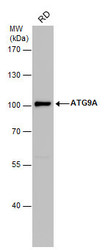
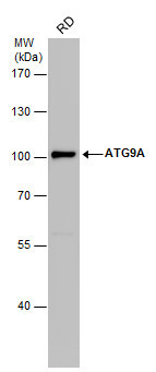
- ATG9A antibody detects ATG9A protein by western blot analysis. Whole cell extracts (30 ?g) was separated by 7.5% SDS-PAGE, and the membrane was blotted with ATG9A antibody (GTX128427) diluted by 1:1000. The HRP-conjugated anti-rabbit IgG antibody (GTX213110-01) was used to detect the primary antibody.
Supportive validation
- Submitted by
- GeneTex (provider)
- Main image
- Experimental details
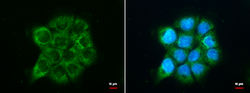
- ATG9A antibody detects ATG9A protein at cytoplasm by immunofluorescent analysis. Sample: A431 cells were fixed in 100% MeOH for 5 min.Green: ATG9A protein stained by ATG9A antibody (GTX128427) diluted at 1:500.Blue: Hoechst 33342 staining.

Supportive validation
- Submitted by
- GeneTex (provider)
- Main image
- Experimental details
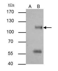
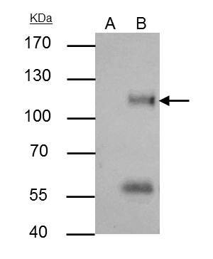
- ATG9A antibody immunoprecipitates ATG9A protein in IP experiments.IP samples: HeLa whole cell extractA. Control with 4 £gg of preimmune Rabbit IgGB. Immunoprecipitation of ATG9A protein by 4 £gg ATG9A antibody (GTX128427)7.5 % SDS-PAGEThe immunoprecipitated ATG9A protein was detected by ATG9A antibody (GTX128427) diluted at 1:500.[EasyBlot anti-rabbit IgG (GTX221666-01) was used as a secondary reagent]
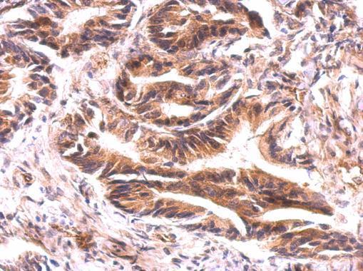
Supportive validation
- Submitted by
- GeneTex (provider)
- Main image

- Experimental details
- ATG9A antibody detects ATG9A protein on colon carcinoma by immunohistochemical analysis. Sample: Paraffin-embedded colon carcinoma. ATG9A antibody (GTX128427) dilution: 1:500.