Explore
 Validate
 Learn
Learn Western blot
 Immunocytochemistry
Antibody data
- Antibody Data
- Antigen structure
- References [6]
- Comments [0]
- Validations
- Immunocytochemistry [1]
- Immunohistochemistry [1]
Submit
Validation data
Reference
Comment
Report error
- Product number
- HPA029159 - Provider product page

- Provider
- Atlas Antibodies
- Proper citation
- Atlas Antibodies Cat#HPA029159, RRID:AB_10599619
- Product name
- Anti-CTNNB1
- Antibody type
- Polyclonal
- Description
- Polyclonal Antibody against Human CTNNB1, Gene description: catenin (cadherin-associated protein), beta 1, 88kDa, Alternative Gene Names: armadillo, beta-catenin, CTNNB, Validated applications: WB, IHC, ICC, Uniprot ID: P35222, Storage: Store at +4°C for short term storage. Long time storage is recommended at -20°C.
- Reactivity
- Human, Mouse, Rat
- Host
- Rabbit
- Conjugate
- Unconjugated
- Isotype
- IgG
- Vial size
- 100 µl
- Concentration
- 0.3 mg/ml
- Storage
- Store at +4°C for short term storage. Long time storage is recommended at -20°C.
- Handling
- The antibody solution should be gently mixed before use.
Submitted references CD142 Identifies Neoplastic Desmoid Tumor Cells, Uncovering Interactions Between Neoplastic and Stromal Cells That Drive Proliferation
Proteogenomic insights suggest druggable pathways in endometrial carcinoma.
Centyrin ligands for extrahepatic delivery of siRNA
The Role of Wnt/β-Catenin Pathway Mediators in Aortic Valve Stenosis
The NFκB subunit RELA is a master transcriptional regulator of the committed epithelial-mesenchymal transition in airway epithelial cells
The Urotensin II System and Carotid Atherosclerosis: A Role in Vascular Calcification
Al-Jazrawe M, Xu S, Poon R, Wei Q, Przybyl J, Varma S, van de Rijn M, Alman B
Cancer Research Communications 2023;3(4):697-708
Cancer Research Communications 2023;3(4):697-708
Proteogenomic insights suggest druggable pathways in endometrial carcinoma.
Dou Y, Katsnelson L, Gritsenko MA, Hu Y, Reva B, Hong R, Wang YT, Kolodziejczak I, Lu RJ, Tsai CF, Bu W, Liu W, Guo X, An E, Arend RC, Bavarva J, Chen L, Chu RK, Czekański A, Davoli T, Demicco EG, DeLair D, Devereaux K, Dhanasekaran SM, Dottino P, Dover B, Fillmore TL, Foxall M, Hermann CE, Hiltke T, Hostetter G, Jędryka M, Jewell SD, Johnson I, Kahn AG, Ku AT, Kumar-Sinha C, Kurzawa P, Lazar AJ, Lazcano R, Lei JT, Li Y, Liao Y, Lih TM, Lin TT, Martignetti JA, Masand RP, Matkowski R, McKerrow W, Mesri M, Monroe ME, Moon J, Moore RJ, Nestor MD, Newton C, Omelchenko T, Omenn GS, Payne SH, Petyuk VA, Robles AI, Rodriguez H, Ruggles KV, Rykunov D, Savage SR, Schepmoes AA, Shi T, Shi Z, Tan J, Taylor M, Thiagarajan M, Wang JM, Weitz KK, Wen B, Williams CM, Wu Y, Wyczalkowski MA, Yi X, Zhang X, Zhao R, Mutch D, Chinnaiyan AM, Smith RD, Nesvizhskii AI, Wang P, Wiznerowicz M, Ding L, Mani DR, Zhang H, Anderson ML, Rodland KD, Zhang B, Liu T, Fenyö D, Clinical Proteomic Tumor Analysis Consortium
Cancer cell 2023 Sep 11;41(9):1586-1605.e15
Cancer cell 2023 Sep 11;41(9):1586-1605.e15
Centyrin ligands for extrahepatic delivery of siRNA
Klein D, Goldberg S, Theile C, Dambra R, Haskell K, Kuhar E, Lin T, Parmar R, Manoharan M, Richter M, Wu M, Mendrola Zarazowski J, Jadhav V, Maier M, Sepp-Lorenzino L, O’Neil K, Dudkin V
Molecular Therapy 2021;29(6):2053-2066
Molecular Therapy 2021;29(6):2053-2066
The Role of Wnt/β-Catenin Pathway Mediators in Aortic Valve Stenosis
Khan K, Yu B, Kiwan C, Shalal Y, Filimon S, Cipro M, Shum-Tim D, Cecere R, Schwertani A
Frontiers in Cell and Developmental Biology 2020;8
Frontiers in Cell and Developmental Biology 2020;8
The NFκB subunit RELA is a master transcriptional regulator of the committed epithelial-mesenchymal transition in airway epithelial cells
Tian B, Widen S, Yang J, Wood T, Kudlicki A, Zhao Y, Brasier A
Journal of Biological Chemistry 2018;293(42):16528-16545
Journal of Biological Chemistry 2018;293(42):16528-16545
The Urotensin II System and Carotid Atherosclerosis: A Role in Vascular Calcification
Albanese I, Daskalopoulou S, Yu B, You Z, Genest J, Alsheikh-Ali A, Schwertani A
Frontiers in Pharmacology 2016;7
Frontiers in Pharmacology 2016;7
No comments: Submit comment
Supportive validation
- Submitted by
- Atlas Antibodies (provider)
- Main image
- Experimental details
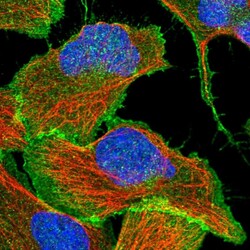
- Immunofluorescent staining of human cell line U-2 OS shows localization to plasma membrane.
- Sample type
- Human
Supportive validation
- Submitted by
- Atlas Antibodies (provider)
- Enhanced method
- Orthogonal validation
- Main image
- Experimental details
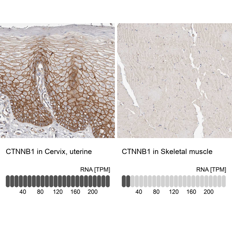
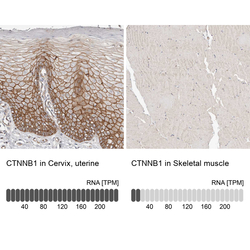
- Immunohistochemistry analysis in human cervix, uterine and skeletal muscle tissues using HPA029159 antibody. Corresponding CTNNB1 RNA-seq data are presented for the same tissues.
- Sample type
- Human
- Protocol
- Protocol