Explore
 Validate
 Learn
Learn Western blot
Antibody data
- Antibody Data
- Antigen structure
- References [1]
- Comments [0]
- Validations
- Western blot [4]
- Immunocytochemistry [1]
- Immunohistochemistry [9]
- Flow cytometry [1]
- Other assay [1]
Submit
Validation data
Reference
Comment
Report error
- Product number
- UM500015 - Provider product page

- Provider
- OriGene
- Proper citation
- OriGene Cat#UM500015, RRID:AB_2629030
- Product name
- Beta-Catenin (CTNNB1) mouse monoclonal antibody, clone UMAB15
- Antibody type
- Monoclonal
- Description
- Beta-Catenin (CTNNB1) mouse monoclonal antibody, clone UMAB15
- Host
- Mouse
- Conjugate
- Unconjugated
- Epitope
- CTNNB1
- Isotype
- IgG
- Antibody clone number
- UMAB15
- Vial size
- 100 µl
- Concentration
- 0.97 mg/ml
Submitted references A Novel Phenotype of Familial Hyperaldosteronism Type III: Concurrence of Aldosteronism and Cushing's Syndrome.
Tong A, Liu G, Wang F, Jiang J, Yan Z, Zhang D, Zhang Y, Cai J
The Journal of clinical endocrinology and metabolism 2016 Nov;101(11):4290-4297
The Journal of clinical endocrinology and metabolism 2016 Nov;101(11):4290-4297
No comments: Submit comment
Supportive validation
- Submitted by
- OriGene (provider)
- Main image
- Experimental details
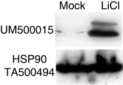
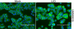
- Western analysis of HEK293T cells without treatment or treated with 30mM LiCl for 16 hours, using anti-beta-catenin antibody (clone UMAB15). Anti-HSP90 (TA500494) was used as internal loading control.
- Validation comment
- WB

- Submitted by
- OriGene (provider)
- Main image
- Experimental details
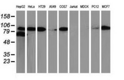
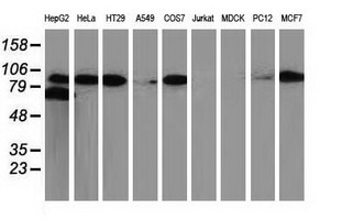
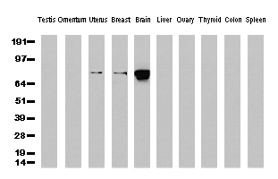
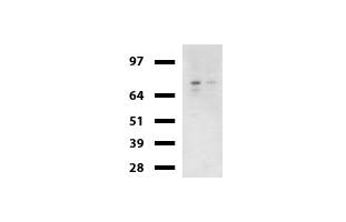
- Western blot analysis of extracts (35ug) from 9 different cell lines by using anti-CTNNB1 monoclonal antibody.
- Validation comment
- WB
- Submitted by
- OriGene (provider)
- Main image

- Experimental details
- Western Blot analysis of 10 different human tissue lysates (10ug) by using Anti-?-Catenin monoclonal antibody (Clone UMAB15, 1:500)
- Validation comment
- WB
- Submitted by
- OriGene (provider)
- Main image
- Experimental details
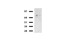
- Western blot of mouse tissue lysates (20ug) from Uterus and Ovary. Primary antibody diluation: 1:500. Secondary antibody dilution: Mouse TrueBlot? Ultra (1:1000).
- Validation comment
- WB
Supportive validation
- Submitted by
- OriGene (provider)
- Main image
- Experimental details
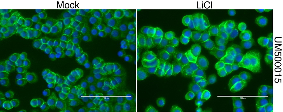
- Immunofluorescent image of HT-29 cells without treatment (left) or treated with 30mM LiCl (right), using anti-beta-catenin antibody (Clone UMAB15). Nuclei were labeled by Hoechst 33242 (blue).
- Validation comment
- IF
Supportive validation
- Submitted by
- OriGene (provider)
- Main image

- Experimental details
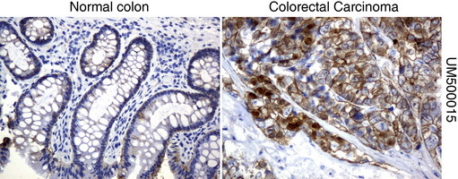
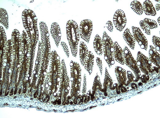
- Immunohistochemical staining of paraffin-embedded Human normal colon and colorectal carcinoma tissues using anti-beta-catenin mouse monoclonal antibody. Note: beta-catenin is mostly located at membrane/cytoplasmic region, but exhibited intensive nuclear s
- Validation comment
- IHC
- Submitted by
- OriGene (provider)
- Main image

- Experimental details
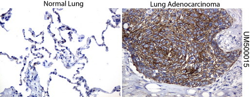
- Immunohistochemical staining of paraffin-embedded human normal lung and lung Adenocarcinoma tissues using anti-beta-catenin mouse monoclonal antibody. (Clone UMAB15, dilution 1:100; heat-induced epitope retrieval by 10mM citric buffer, pH6.0, 120C for 3mi
- Validation comment
- IHC
- Submitted by
- OriGene (provider)
- Main image
- Experimental details
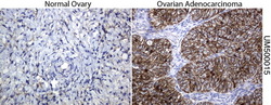
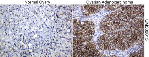
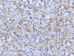
- Immunohistochemical staining of paraffin-embedded human normal ovary and Adenocarcinoma of the ovary tissues using anti-beta-catenin mouse monoclonal antibody. (Clone UMAB15, dilution 1:100; heat-induced epitope retrieval by 10mM citric buffer, pH6.0, 120
- Validation comment
- IHC
- Submitted by
- OriGene (provider)
- Main image
- Experimental details
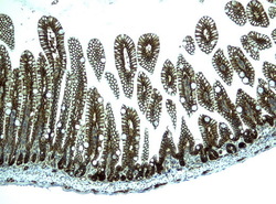
- Immunohistochemical staining of paraffin-embedded Mouse colon tissue using anti-beta-catenin mouse monoclonal antibody. (Clone UMAB15, dilution 1:100; heat-induced epitope retrieval by 10mM citric buffer, pH6.0, 120C for 3min)
- Validation comment
- IHC
- Submitted by
- OriGene (provider)
- Main image
- Experimental details
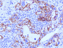
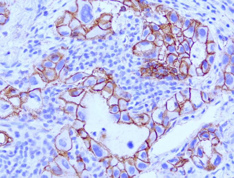
- Immunohistochemical staining of paraffin-embedded human lung cancer with mouse anti-Beta Catenin clone UMAB15 1:200 using HIER citrate pressure chamber. Tumor cells show positive membrane staining.
- Validation comment
- IHC
- Submitted by
- OriGene (provider)
- Main image
- Experimental details
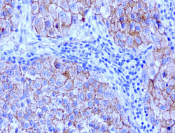
- Immunohistochemical staining of paraffin-embedded human melanoma metastisis to lymph node with mouse anti-Beta Catenin clone UMAB15 1:200 using HIER citrate pressure chamber. Tumor cells show positive membrane and cytoplasmic staining.
- Validation comment
- IHC
- Submitted by
- OriGene (provider)
- Main image
- Experimental details
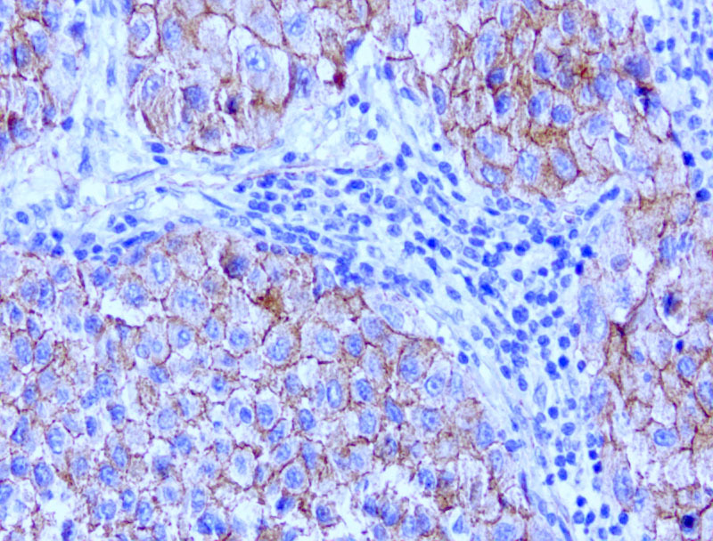
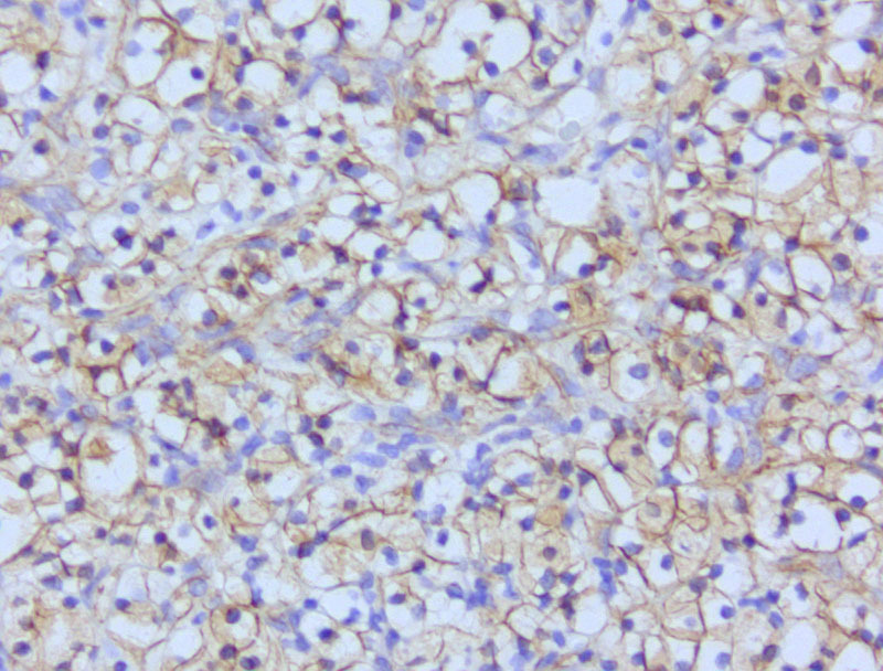
- Immunohistochemical staining of paraffin-embedded human renal cell carcinoma with mouse anti-Beta Catenin clone UMAB15 1:200 using HIER citrate pressure chamber. Tumor cells show positive membrane staining.
- Validation comment
- IHC
- Submitted by
- OriGene (provider)
- Main image
- Experimental details
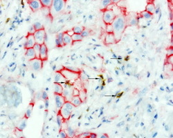
- Sequential double staining of paraffin human melanoma using bCatenin UM500015 (red) and PD1 UM800091 (brown). Both abs at 1:800 dilution of 1mg/mL. Anti-PD1: heat-induced epitope retrieval with Accel; anti-bCatenin: citrate pH6.0. Image shows the tumor cells are strongly positve for b-catenin (red) and negative for PD1. The arrows point to the activated T cells (brown) show strong membranous and cytoplasmic staining of PD1 and no staining with bCatenin.
- Validation comment
- IHC

- Submitted by
- OriGene (provider)
- Main image
- Experimental details
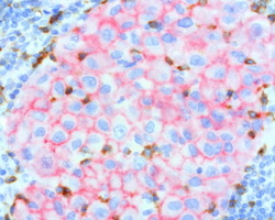
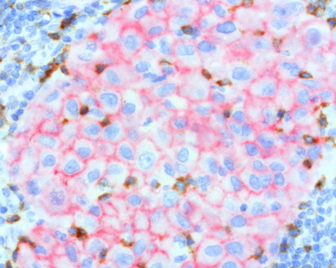
- Sequential double staining of paraffin human melanoma using bCatenin UM500015 (red) and PD1 UM800091 (brown). Both abs at 1:800 dilution of 1mg/mL. Anti-PD1: heat-induced epitope retrieval with Accel; anti-bCatenin: citrate pH6.0. The image of shows the tumor cells are strongly positve for beta-catenin (red) are negative for PD-1. The activated TCells (brown) show strong membranous and cytoplasmic staining for PD-1 and no staining with Beta Catenin.
- Validation comment
- IHC
Supportive validation
- Submitted by
- OriGene (provider)
- Main image

- Experimental details
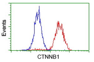
- Flow cytometric Analysis of Hela cells, using anti-CTNNB1 antibody(UM500015),(Red), compared to a nonspecific negative control antibody,(Blue).
- Validation comment
- FC
Supportive validation
- Submitted by
- OriGene (provider)
- Main image
- Experimental details
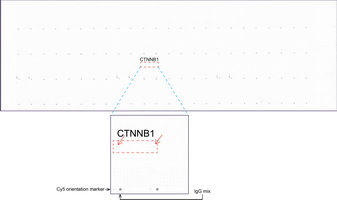
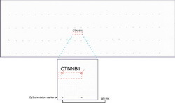
- OriGene overexpression protein microarray chip was immunostained with UltraMAB anti-beta-catenin mouse monoclonal antibody (Clone UMAB15). The positive reactive proteins are highlighted with two red arrows in the enlarged subarray. All the positive controls spotted in this subarray are also labeled for clarification. These data show that UltraMAB anti-beta-catenin (Clone UMAB15) very specifically recognizes beta-catenin antigen on OriGene protein microarray chip.
- Validation comment
- 10K-CHIP