Explore
 Validate
 Learn
Learn Western blot
Antibody data
- Antibody Data
- Antigen structure
- References [0]
- Comments [0]
- Validations
- Western blot [1]
- ELISA [1]
- Immunocytochemistry [2]
- Proximity ligation assay [2]
Submit
Validation data
Reference
Comment
Report error
- Product number
- H00001499-M02 - Provider product page

- Provider
- Abnova Corporation
- Proper citation
- Abnova Corporation Cat#H00001499-M02, RRID:AB_875480
- Product name
- CTNNB1 monoclonal antibody (M02), clone 1C9
- Antibody type
- Monoclonal
- Description
- Mouse monoclonal antibody raised against a partial recombinant CTNNB1.
- Antigen sequence
LFRTEPMAWNETADLGLDIGAQGEPLGYRQDDPSY
RSFHSGGYGQDALGMDPMMEHEMGGHHPGADYPVD
GLPDLGHAQDLMDGLPPGDSNQLAWFDTDL- Isotype
- IgG
- Antibody clone number
- 1C9
- Storage
- Store at -20°C or lower. Aliquot to avoid repeated freezing and thawing.
No comments: Submit comment
Supportive validation
- Submitted by
- Abnova Corporation (provider)
- Main image
- Experimental details
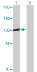
- Western Blot analysis of CTNNB1 expression in transfected 293T cell line by CTNNB1 monoclonal antibody (M02), clone 1C9.Lane 1: CTNNB1 transfected lysate(85.5 KDa).Lane 2: Non-transfected lysate.

Supportive validation
- Submitted by
- Abnova Corporation (provider)
- Main image
- Experimental details
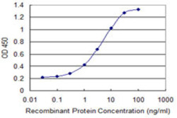
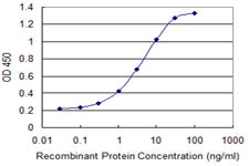
- Detection limit for recombinant GST tagged CTNNB1 is 0.1 ng/ml as a capture antibody.
- Validation comment
- Sandwich ELISA (Recombinant protein)
- Protocol
- Protocol
Supportive validation
- Submitted by
- Abnova Corporation (provider)
- Main image

- Experimental details
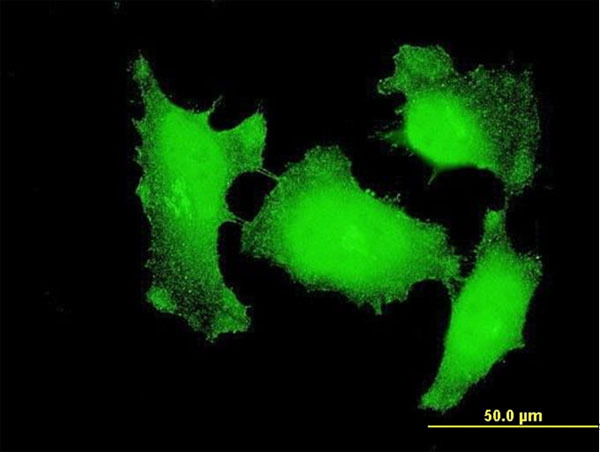
- Immunofluorescence of monoclonal antibody to CTNNB1 on HeLa cell. [antibody concentration 10 ug/ml]
- Validation comment
- Immunofluorescence
- Protocol
- Protocol
- Submitted by
- Abnova Corporation (provider)
- Main image
- Experimental details
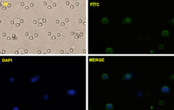
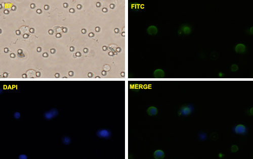
- PC-3 MM2 cells were stained with CTNNB1-FITC labeled monoclonal antibody (Green). The cell nucleus were counterstained with DAPI (Blue).
- Validation comment
- Immunofluorescence (Circulating Tumor Cell)
Supportive validation
- Submitted by
- Abnova Corporation (provider)
- Main image
- Experimental details
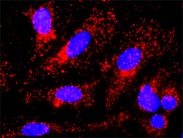
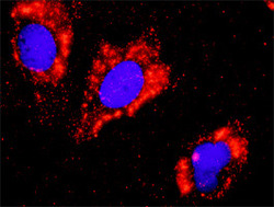
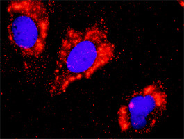
- Proximity Ligation Analysis of protein-protein interactions between FLT1 and CTNNB1. Huh7 cells were stained with anti-FLT1 rabbit purified polyclonal 1:1200 and anti-CTNNB1 mouse monoclonal antibody 1:50. Each red dot represents the detection of protein-protein interaction complex, and nuclei were counterstained with DAPI (blue).
- Validation comment
- In situ Proximity Ligation Assay (Cell)
- Submitted by
- Abnova Corporation (provider)
- Main image

- Experimental details
- Proximity Ligation Analysis of protein-protein interactions between GSK3B and CTNNB1. HeLa cells were stained with anti-GSK3B rabbit purified polyclonal 1:1200 and anti-CTNNB1 mouse monoclonal antibody 1:50. Each red dot represents the detection of protein-protein interaction complex, and nuclei were counterstained with DAPI (blue).
- Validation comment
- In situ Proximity Ligation Assay (Cell)