Explore
 Validate
 Learn
Learn Flow cytometry
Antibody data
- Antibody Data
- Antigen structure
- References [1]
- Comments [0]
- Validations
- Flow cytometry [1]
- Other assay [1]
Submit
Validation data
Reference
Comment
Report error
- Product number
- 12-2567-42 - Provider product page

- Provider
- Invitrogen Antibodies
- Product name
- beta Catenin Monoclonal Antibody (15B8), PE, eBioscience™
- Antibody type
- Monoclonal
- Antigen
- Other
- Description
- Description: The 15B8 monoclonal antibody reacts with human and mouse beta-catenin, one member of a family of catenins, which are intracellular proteins that interact with cadherins to mediate cellular adhesion. More specifically, beta-catenin binds to the cytoplasmic tail of E-cadherin. In addition, this molecule is a component of the canonical Wnt signaling pathway. In the absence of Wnt binding its receptor, beta-catenin is phosphorylated and resides in the cytoplasm where it is eventually targeted for degradation by ubiquitination. Upon Wnt binding, beta-catenin becomes dephosphorylated, translocates to the nucleus, and modulates gene expression in partnership with the transcription factors T cell factor (TCF) and lymphocyte enhancer binding factor (LEF). Expression of beta-catenin is found in a wide variety of non-immune and immune tissues, including thymocytes and T and B lymphocytes. The Wnt and beta-catenin signaling pathway has been demonstrated to play a crucial role in the development of T, B, and hematopoietic stem cells. Applications Reported: This 15B8 antibody has been reported for use in intracellular staining followed by flow cytometric analysis. Applications Tested: This 15B8 antibody has been pre-titrated and tested by intracellular staining and flow cytometric analysis of Jurkat cell line using the Foxp3/Transcription Factor Staining Buffer Set (Product # 00-5523-00) and protocol. This can be used at 5 µL (0.125 µg) per test. A test is defined as the amount (µg) of antibody that will stain a cell sample in a final volume of 100 µL. Cell number should be determined empirically but can range from 10^5 to 10^8 cells/test. Excitation: 488-561 nm; Emission: 578 nm; Laser: Blue Laser, Green Laser, Yellow-Green Laser. Filtration: 0.2 µm post-manufacturing filtered.
- Reactivity
- Human, Mouse
- Host
- Mouse
- Conjugate
- Yellow dye
- Isotype
- IgG
- Antibody clone number
- 15B8
- Vial size
- 100 Tests
- Concentration
- 5 µL/Test
- Storage
- 4° C, store in dark, DO NOT FREEZE!
Submitted references Wnt activation promotes memory T cell polyfunctionality via epigenetic regulator PRMT1.
Sung BY, Lin YH, Kong Q, Shah PD, Glick Bieler J, Palmer S, Weinhold KJ, Chang HR, Huang H, Avery RK, Schneck J, Chiu YL
The Journal of clinical investigation 2022 Jan 18;132(2)
The Journal of clinical investigation 2022 Jan 18;132(2)
No comments: Submit comment
Supportive validation
- Submitted by
- Invitrogen Antibodies (provider)
- Main image
- Experimental details
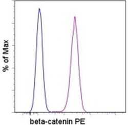
- Staining of Jurkat cell line with Mouse IgG1 K Isotype Control PE (Product # 12-4714-81) (blue histogram) or Anti-Human/Mouse beta-Catenin PE (purple histogram). Total viable cells were used for analysis.
- Conjugate
- Yellow dye

Supportive validation
- Submitted by
- Invitrogen Antibodies (provider)
- Main image

- Experimental details
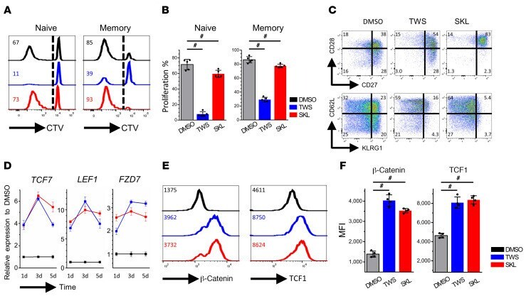
- Figure 1 Wnt signaling alters human memory CD8 + T cell proliferation and differentiation. ( A ) FACS-sorted human naive and memory CD8 + T cells were stained with CTV and stimulated with CD3/CD28 in DMSO (black), TWS119 (blue), and SKL2001 (red) for 7 days. Numbers represent the percentages of divided cells in each treatment. ( B ) Average proliferative response for each treatment ( n = 5). Dunn's test for multiple comparisons. ( C ) Memory CD8 + T cells in different treatments for 7 days were stained with phenotypic markers. Numbers represent the percentages of cells in each quadrant. ( D ) Quantitative RT-PCR analysis of Tcf7, Lef1, and Fzd7 in memory CD8 + T cells with or without Wnt agonist treatment on days 1, 3, and 5. Numbers are normalized to marker expression of the DMSO group at each corresponding time point. ( E ) Memory CD8 + T cells were stimulated with CD3/CD28 and treated with or without Wnt agonists for 7 days. beta-Catenin and TCF1 levels were analyzed by flow cytometry. Numbers represent the MFI of each protein. ( F ) Average MFI of beta-catenin and TCF1 in different treatment conditions ( n = 4). Dunn's test for multiple comparisons. # P < 0.05.
- Conjugate
- Yellow dye