Explore
 Validate
 Learn
Learn Western blot
 Immunoprecipitation
 Immunohistochemistry
Antibody data
- Antibody Data
- Antigen structure
- References [9]
- Comments [0]
- Validations
- Western blot [2]
Submit
Validation data
Reference
Comment
Report error
- Product number
- 14-2567-80 - Provider product page

- Provider
- Invitrogen Antibodies
- Product name
- Anti-beta Catenin Monoclonal Antibody (15B8), eBioscience™
- Antibody type
- Monoclonal
- Antigen
- Other
- Description
- Description: The 15B8 monoclonal antibody reacts with human and mouse beta-catenin, one member of a family of catenins, which are intracellular proteins that interact with cadherins to mediate cellular adhesion. More specifically, beta-catenin binds to the cytoplasmic tail of E-cadherin. In addition, this molecule is a component of the canonical Wnt signaling pathway. In the absence of Wnt binding its receptor, beta-catenin is phosphorylated and resides in the cytoplasm where it is eventually targeted for degradation by ubiquitination. Upon Wnt binding, beta-catenin becomes dephosphorylated, translocates to the nucleus, and modulates gene expression in partnership with the transcription factors T cell factor (TCF) and lymphocyte enhancer binding factor (LEF). Expression of beta-catenin is found in a wide variety of non-immune and immune tissues, including thymocytes and T and B lymphocytes. The Wnt and beta-catenin signaling pathway has been demonstrated to play a crucial role in the development of T, B, and hematopoietic stem cells. Applications Reported: This 15B8 antibody has been reported for use in intracellular staining followed by flow cytometric analysis, immunoprecipitation, western blotting, and immunohistochemical staining of formalin-fixed paraffin embedded tissue sections. (Fluorochrome conjugated 15B8 is recommended for use in intracellular flow cytometry.). Applications Tested: This 15B8 antibody has been tested by western blot analysis on reduced cell lysates prepared from the Jurkat cell line. This antibody can be used at 1-5 µg/mL. This antibody has been tested by immunohistchemistry of formalin-fixed paraffin embedded human tissue using low or high pH antigen retrieval and can be used at less than or equal to 5 µg/mL. It is recommended that the antibody be carefully titrated for optimal performance in the assay of interest. Purity: Greater than 90%, as determined by SDS-PAGE. Aggregation: Less than 10%, as determined by HPLC. Filtration: 0.2 µm post-manufacturing filtered.
- Reactivity
- Human, Mouse
- Host
- Mouse
- Isotype
- IgG
- Antibody clone number
- 15B8
- Vial size
- 25 µg
- Concentration
- 0.5 mg/mL
- Storage
- 4° C
Submitted references Trypanosoma cruzi Exploits Wnt Signaling Pathway to Promote Its Intracellular Replication in Macrophages.
Engineered Microvasculature in PDMS Networks Using Endothelial Cells Derived from Human Induced Pluripotent Stem Cells.
Blocking the epithelial-to-mesenchymal transition pathway abrogates resistance to anti-folate chemotherapy in lung cancer.
Modulation of N-glycosylation by mesalamine facilitates membranous E-cadherin expression in colon epithelial cells.
Expression of intercellular adhesion molecule 1 by hepatocellular carcinoma stem cells and circulating tumor cells.
Sustained expression of pre-TCR induced beta-catenin in post-beta-selection thymocytes blocks T cell development.
The canonical Wnt signaling pathway plays an important role in lymphopoiesis and hematopoiesis.
Deletion of beta-catenin impairs T cell development.
Beta-catenin mediates the interaction of the cadherin-catenin complex with epidermal growth factor receptor.
Volpini X, Ambrosio LF, Fozzatti L, Insfran C, Stempin CC, Cervi L, Motran CC
Frontiers in immunology 2018;9:859
Frontiers in immunology 2018;9:859
Engineered Microvasculature in PDMS Networks Using Endothelial Cells Derived from Human Induced Pluripotent Stem Cells.
Sivarapatna A, Ghaedi M, Xiao Y, Han E, Aryal B, Zhou J, Fernandez-Hernando C, Qyang Y, Hirschi KK, Niklason LE
Cell transplantation 2017 Aug;26(8):1365-1379
Cell transplantation 2017 Aug;26(8):1365-1379
Blocking the epithelial-to-mesenchymal transition pathway abrogates resistance to anti-folate chemotherapy in lung cancer.
Liang SQ, Marti TM, Dorn P, Froment L, Hall SR, Berezowska S, Kocher G, Schmid RA, Peng RW
Cell death & disease 2015 Jul 16;6:e1824
Cell death & disease 2015 Jul 16;6:e1824
Modulation of N-glycosylation by mesalamine facilitates membranous E-cadherin expression in colon epithelial cells.
Khare V, Lang M, Dammann K, Campregher C, Lyakhovich A, Gasche C
Biochemical pharmacology 2014 Jan 15;87(2):312-20
Biochemical pharmacology 2014 Jan 15;87(2):312-20
Expression of intercellular adhesion molecule 1 by hepatocellular carcinoma stem cells and circulating tumor cells.
Liu S, Li N, Yu X, Xiao X, Cheng K, Hu J, Wang J, Zhang D, Cheng S, Liu S
Gastroenterology 2013 May;144(5):1031-1041.e10
Gastroenterology 2013 May;144(5):1031-1041.e10
Sustained expression of pre-TCR induced beta-catenin in post-beta-selection thymocytes blocks T cell development.
Xu M, Sharma A, Hossain MZ, Wiest DL, Sen JM
Journal of immunology (Baltimore, Md. : 1950) 2009 Jan 15;182(2):759-65
Journal of immunology (Baltimore, Md. : 1950) 2009 Jan 15;182(2):759-65
The canonical Wnt signaling pathway plays an important role in lymphopoiesis and hematopoiesis.
Staal FJ, Sen JM
European journal of immunology 2008 Jul;38(7):1788-94
European journal of immunology 2008 Jul;38(7):1788-94
Deletion of beta-catenin impairs T cell development.
Xu Y, Banerjee D, Huelsken J, Birchmeier W, Sen JM
Nature immunology 2003 Dec;4(12):1177-82
Nature immunology 2003 Dec;4(12):1177-82
Beta-catenin mediates the interaction of the cadherin-catenin complex with epidermal growth factor receptor.
Hoschuetzky H, Aberle H, Kemler R
The Journal of cell biology 1994 Dec;127(5):1375-80
The Journal of cell biology 1994 Dec;127(5):1375-80
No comments: Submit comment
Supportive validation
- Submitted by
- Invitrogen Antibodies (provider)
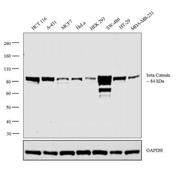
- Main image
- Experimental details
- Western blot analysis was performed on whole cell extracts (30 µg lysate) of HCT 116 (Lane 1), A-431 (Lane 2), MCF7 (Lane 3), HeLa (Lane 4), HEK 293 (Lane 5), SW-480 (Lane 6), HT-29 (Lane 7) and MDA-MB-231 (Lane 8). The blot was probed with beta Catenin Monoclonal Antibody (Product # 14-2567-80, 1:250 dilution) and detected by chemiluminescence using Goat anti-Mouse IgG (H+L) Superclonal™ Secondary Antibody, HRP conjugate (Product # A28177, 0.25 µg/mL, 1:4000 dilution). A 85 kDa band corresponding to beta Catenin was observed across the cell lines tested.
- Submitted by
- Invitrogen Antibodies (provider)
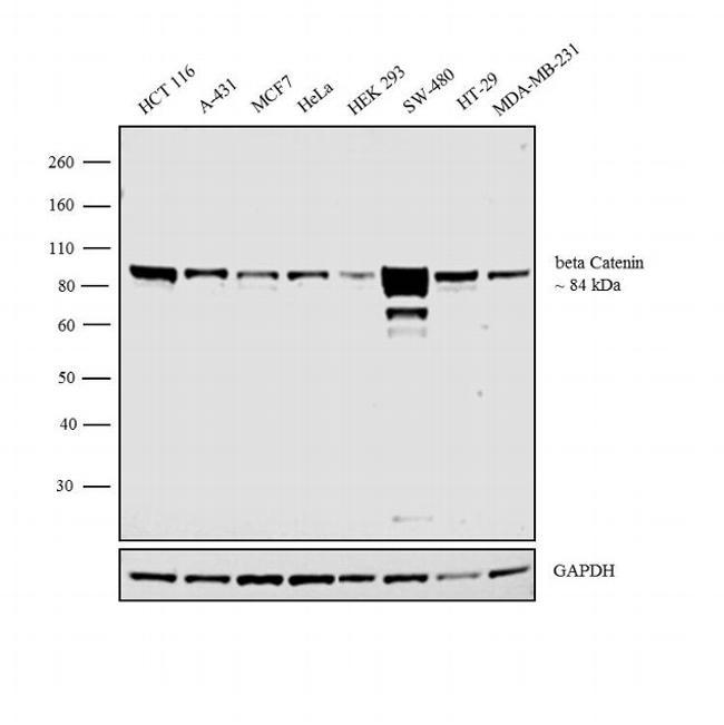
- Main image

- Experimental details
- Western blot analysis was performed on whole cell extracts (30 µg lysate) of HCT 116 (Lane 1), A-431 (Lane 2), MCF7 (Lane 3), HeLa (Lane 4), HEK 293 (Lane 5), SW-480 (Lane 6), HT-29 (Lane 7) and MDA-MB-231 (Lane 8). The blot was probed with beta Catenin Monoclonal Antibody (Product # 14-2567-82, 2µg/mL) and detected by chemiluminescence using Goat anti-Mouse IgG (H+L) Superclonal™ Secondary Antibody, HRP conjugate (Product # A28177, 0.25 µg/mL, 1:4000 dilution). A 85 kDa band corresponding to beta Catenin was observed across the cell lines tested.