 Explore
 Validate
 Learn
Learn Western blot
 Immunocytochemistry
Antibody data
- Antibody Data
- Antigen structure
- References [12]
- Comments [0]
- Validations
- Western blot [5]
- Immunohistochemistry [4]
- Flow cytometry [2]
Submit
Validation data
Reference
Comment
Report error
- Product number
- NBP1-54467 - Provider product page

- Provider
- Novus Biologicals
- Proper citation
- Novus Cat#NBP1-54467, RRID:AB_11006210
- Product name
- Mouse Monoclonal beta-Catenin Antibody
- Antibody type
- Monoclonal
- Description
- Protein G purified.
- Reactivity
- Human, Mouse, Rat, Chicken/Avian, Simian
- Host
- Mouse
- Isotype
- IgG
- Vial size
- 0.1 ml
- Concentration
- 1.0 mg/ml
- Storage
- Aliquot and store at -20C or -80C. Avoid freeze-thaw cycles.
Submitted references Expression of a Degradation-Resistant β-Catenin Mutant in Osteocytes Protects the Skeleton From Mechanodeprivation-Induced Bone Wasting.
Anti-metastatic effects of isolinderalactone via the inhibition of MMP-2 and up regulation of NM23-H1 expression in human lung cancer A549 cells.
Physical disruption of cell-cell contact induces VEGF expression in RPE cells.
Haploinsufficiency of RCBTB1 is associated with Coats disease and familial exudative vitreoretinopathy.
Canonical Wnt signaling activity in early stages of chick lung development.
Characterization of the interactions of alpha-catenin with alpha-actinin and beta-catenin/plakoglobin.
Expression of N-cadherin by human squamous carcinoma cells induces a scattered fibroblastic phenotype with disrupted cell-cell adhesion.
Expression of N-cadherin by human squamous carcinoma cells induces a scattered fibroblastic phenotype with disrupted cell-cell adhesion.
Interaction of alpha-actinin with the cadherin/catenin cell-cell adhesion complex via alpha-catenin.
Interaction of alpha-actinin with the cadherin/catenin cell-cell adhesion complex via alpha-catenin.
Identification of plakoglobin domains required for association with N-cadherin and alpha-catenin.
Identification of plakoglobin domains required for association with N-cadherin and alpha-catenin.
Bullock WA, Hoggatt AM, Horan DJ, Lewis KJ, Yokota H, Hann S, Warman ML, Sebastian A, Loots GG, Pavalko FM, Robling AG
Journal of bone and mineral research : the official journal of the American Society for Bone and Mineral Research 2019 Oct;34(10):1964-1975
Journal of bone and mineral research : the official journal of the American Society for Bone and Mineral Research 2019 Oct;34(10):1964-1975
Anti-metastatic effects of isolinderalactone via the inhibition of MMP-2 and up regulation of NM23-H1 expression in human lung cancer A549 cells.
Chuang CH, Wang LY, Wong YM, Lin ES
Oncology letters 2018 Apr;15(4):4690-4696
Oncology letters 2018 Apr;15(4):4690-4696
Physical disruption of cell-cell contact induces VEGF expression in RPE cells.
Farjood F, Vargis E
Molecular vision 2017;23:431-446
Molecular vision 2017;23:431-446
Haploinsufficiency of RCBTB1 is associated with Coats disease and familial exudative vitreoretinopathy.
Wu JH, Liu JH, Ko YC, Wang CT, Chung YC, Chu KC, Liu TT, Chao HM, Jiang YJ, Chen SJ, Chung MY
Human molecular genetics 2016 Apr 15;25(8):1637-47
Human molecular genetics 2016 Apr 15;25(8):1637-47
Canonical Wnt signaling activity in early stages of chick lung development.
Moura RS, Carvalho-Correia E, daMota P, Correia-Pinto J
PloS one 2014;9(12):e112388
PloS one 2014;9(12):e112388
Characterization of the interactions of alpha-catenin with alpha-actinin and beta-catenin/plakoglobin.
Nieset JE, Redfield AR, Jin F, Knudsen KA, Johnson KR, Wheelock MJ
Journal of cell science 1997 Apr;110 ( Pt 8):1013-22
Journal of cell science 1997 Apr;110 ( Pt 8):1013-22
Expression of N-cadherin by human squamous carcinoma cells induces a scattered fibroblastic phenotype with disrupted cell-cell adhesion.
Islam S, Carey TE, Wolf GT, Wheelock MJ, Johnson KR
The Journal of cell biology 1996 Dec;135(6 Pt 1):1643-54
The Journal of cell biology 1996 Dec;135(6 Pt 1):1643-54
Interaction of alpha-actinin with the cadherin/catenin cell-cell adhesion complex via alpha-catenin.
Knudsen KA, Soler AP, Johnson KR, Wheelock MJ
The Journal of cell biology 1995 Jul;130(1):67-77
The Journal of cell biology 1995 Jul;130(1):67-77
Interaction of alpha-actinin with the cadherin/catenin cell-cell adhesion complex via alpha-catenin.
Knudsen KA, Soler AP, Johnson KR, Wheelock MJ
The Journal of cell biology 1995 Jul;130(1):67-77
The Journal of cell biology 1995 Jul;130(1):67-77
Identification of plakoglobin domains required for association with N-cadherin and alpha-catenin.
Sacco PA, McGranahan TM, Wheelock MJ, Johnson KR
The Journal of biological chemistry 1995 Aug 25;270(34):20201-6
The Journal of biological chemistry 1995 Aug 25;270(34):20201-6
Identification of plakoglobin domains required for association with N-cadherin and alpha-catenin.
Sacco PA, McGranahan TM, Wheelock MJ, Johnson KR
The Journal of biological chemistry 1995 Aug 25;270(34):20201-6
The Journal of biological chemistry 1995 Aug 25;270(34):20201-6
No comments: Submit comment
Supportive validation
- Submitted by
- Novus Biologicals (provider)
- Main image
- Experimental details
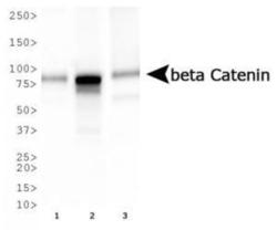
- Western Blot: beta-Catenin Antibody (12F7) [NBP1-54467] - Analysis of beta Catenin expression in 1) HepG2, 2) MCF7, and 3) Cos7 whole cell lysates using NBP1-54467.
- Submitted by
- Novus Biologicals (provider)
- Main image

- Experimental details
- Simple Western: beta-Catenin Antibody (12F7) [NBP1-54467] - Simple Western lane view shows a specific band for Beta- Catenin in 0.5 mg/ml of HepG2 lysate. This experiment was performed under reducing conditions using the 12-230 kDa separation system.

- Submitted by
- Novus Biologicals (provider)
- Main image

- Experimental details
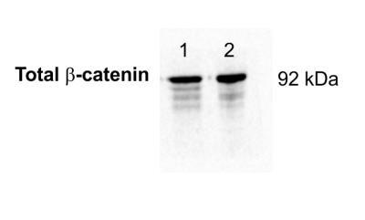
- Western Blot: beta-Catenin Antibody (12F7) [NBP1-54467] - Analysis of beta- Catenin in embryonic lung (lane 1) and embryonic limb (lane 2) lysates using anti-beta- Catenin antibody. Each lane was loaded with 5ug of protein sample. Image from verified customer review.
- Submitted by
- Novus Biologicals (provider)
- Main image

- Experimental details
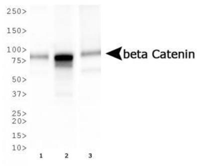
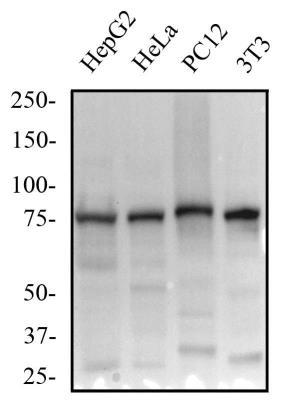
- Western Blot: beta-Catenin Antibody (12F7) [NBP1-54467] - Total protein from human HepG2 and HeLa cells, rat PC12 cells and mouse 3T3 cells was separated on a 7.5% gel by SDS-PAGE, transferred to PVDF membrane and blocked in 5% non-fat milk in TBST. The membrane was probed with 1.0 ug/ml anti-Beta catenin in 1% non-fat milk in TBST and detected with an anti-mouse HRP secondary antibody using chemiluminescence.
- Submitted by
- Novus Biologicals (provider)
- Main image

- Experimental details
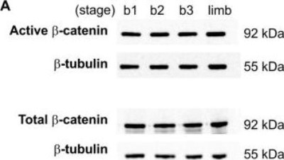
- Western Blot: beta-Catenin Antibody (12F7) [NBP1-54467] - Activity of Wnt/beta-catenin pathway in the embryonic chick lung. Western blot analysis of active and total beta-catenin in stage b1, b2 and b3 lungs, and stage 24 limb (as positive control). Control loading was performed using beta-tubulin (55 kDa). Total and active beta-catenin correspond to 92 kDa. Image collected and cropped by CiteAb from the following publication (http://dx.plos.org/10.1371/journal.pone.0112388), licensed under a CC-BY licence.
Supportive validation
- Submitted by
- Novus Biologicals (provider)
- Main image

- Experimental details
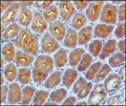
- Immunohistochemistry-Paraffin: beta-Catenin Antibody (12F7) [NBP1-54467] - Analysis of beta Catenin in mouse intestine using DAB with hematoxylin counterstain.

- Submitted by
- Novus Biologicals (provider)
- Main image
- Experimental details
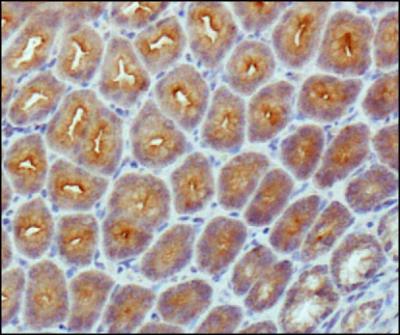
- Immunohistochemistry-Paraffin: beta-Catenin Antibody (12F7) [NBP1-54467] - Analysis of beta- Catenin in mouse intestine using DAB with hematoxylin counterstain.
- Submitted by
- Novus Biologicals (provider)
- Main image

- Experimental details
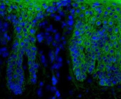
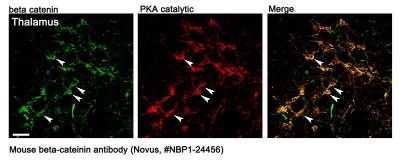
- Immunohistochemistry: beta-Catenin Antibody (12F7) [NBP1-54467] - Beta-catening (green) was detected in human skin (psoriasis) using beta-catenin-FITC antibody (1:40) in PBS for 1 hour. Nuclei were stained with Dapi (blue). Image from verified customer review. Image using the FITC format of this antibody.
- Submitted by
- Novus Biologicals (provider)
- Main image

- Experimental details
- Immunohistochemistry: beta-Catenin Antibody (12F7) [NBP1-54467] - IF beta-Catenin staining of mouse brain tissue. This image submitted by a verified customer review.
Supportive validation
- Submitted by
- Novus Biologicals (provider)
- Main image
- Experimental details
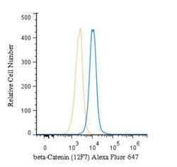
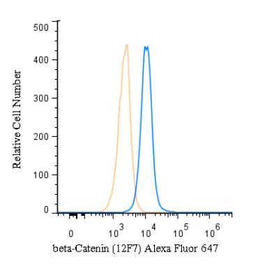
- Flow (Intracellular): beta-Catenin Antibody (12F7) [NBP1-54467] - An intracellular stain was performed on HeLa cells with beta-Catenin Antibody (12F7) NBP1-54467AF647 (blue) and a matched isotype control (orange). Cells were fixed with 4% PFA and then permeabilized with 0.1% saponin. Cells were incubated in an antibody dilution of 5 ug/mL for 30 minutes at room temperature. Both antibodies were conjugated to Alexa Fluor 647.
- Submitted by
- Novus Biologicals (provider)
- Main image

- Experimental details
- Flow Cytometry: beta-Catenin Antibody (12F7) [NBP1-54467] - An intracellular stain was performed on SK-MEL-28 cells with beta-Catenin Antibody [12F7] NBP1-54467B (blue) and a matched isotype control (orange). Both antibodies were conjugated to Biotin. Cells were fixed with 4% PFA and then permeabilized with 0.1% saponin. Cells were incubated in an antibody dilution of 2.5 ug/mL for 30 minutes at room temperature, followed by Streptavidin - R-Phycoerythrin Protein (2012-1000, Novus Biologicals).
