 Explore
 Validate
 Learn
Learn Western blot
Antibody data
- Antibody Data
- Antigen structure
- References [4]
- Comments [0]
- Validations
- Western blot [3]
- Immunocytochemistry [3]
- Immunohistochemistry [2]
- Other assay [2]
Submit
Validation data
Reference
Comment
Report error
- Product number
- PA5-16762 - Provider product page

- Provider
- Invitrogen Antibodies
- Product name
- beta Catenin Polyclonal Antibody
- Antibody type
- Polyclonal
- Antigen
- Synthetic peptide
- Description
- This antibody reacts with the C-terminal portion of beta-Catenin.
- Concentration
- 0.07 mg/mL
Submitted references LncRNA MNX1-AS1 Contributes to Laryngeal Squamous Cell Carcinoma Growth and Migration by Regulating mir-744-5p/bcl9/β-Catenin Axis.
Persistent Cyfip1 Expression Is Required to Maintain the Adult Subventricular Zone Neurogenic Niche.
Polarized AAVR expression determines infectivity by AAV gene therapy vectors.
Inhibition of discoidin domain receptor 2-mediated lung cancer cells progression by gold nanoparticle-aptamer-assisted delivery of peptides containing transmembrane-juxtamembrane 1/2 domain.
Ma B, Ren G, Xu J, Yin C, Shi Y
Cell transplantation 2021 Jan-Dec;30:9636897211005682
Cell transplantation 2021 Jan-Dec;30:9636897211005682
Persistent Cyfip1 Expression Is Required to Maintain the Adult Subventricular Zone Neurogenic Niche.
Habela CW, Yoon KJ, Kim NS, Taga A, Bell K, Bergles DE, Maragakis NJ, Ming GL, Song H
The Journal of neuroscience : the official journal of the Society for Neuroscience 2020 Mar 4;40(10):2015-2024
The Journal of neuroscience : the official journal of the Society for Neuroscience 2020 Mar 4;40(10):2015-2024
Polarized AAVR expression determines infectivity by AAV gene therapy vectors.
Hamilton BA, Li X, Pezzulo AA, Abou Alaiwa MH, Zabner J
Gene therapy 2019 Jun;26(6):240-249
Gene therapy 2019 Jun;26(6):240-249
Inhibition of discoidin domain receptor 2-mediated lung cancer cells progression by gold nanoparticle-aptamer-assisted delivery of peptides containing transmembrane-juxtamembrane 1/2 domain.
Kim D, Yeom JH, Lee B, Lee K, Bae J, Rhee S
Biochemical and biophysical research communications 2015 Aug 21;464(2):392-5
Biochemical and biophysical research communications 2015 Aug 21;464(2):392-5
No comments: Submit comment
Supportive validation
- Submitted by
- Invitrogen Antibodies (provider)
- Main image

- Experimental details
- Western blot of Catenin beta using Catenin beta Polyclonal Antibody (Product # PA5-16762) on MCF-7 Cells.

- Submitted by
- Invitrogen Antibodies (provider)
- Main image

- Experimental details
- Knockdown of beta Catenin was achieved by transfecting HeLa cells with beta Catenin specific siRNAs (Silencer® select Product # s438 ). Western blot analysis (Fig a) was performed using Membrane extract from the beta Catenin knock down cells (lane 3), non-specific scrambled siRNA transfected cells (lane 2) and untransfected cells (lane 1). The blots were probed with Anti-beta Catenin rabbit Polyclonal Antibody (Product # PA5-16762, 0.2 µg/mL) and Goat anti-Rabbit IgG (H+L) Superclonal™ Secondary Antibody, HRP conjugate (Product # A27036, 0.25 µg/mL, 1:4000 dilution). Densitometric analysis of this western blot is shown in histogram (Fig b). Loss of signal upon siRNA mediated knock down confirms that antibody is specific to beta Catenin.

- Submitted by
- Invitrogen Antibodies (provider)
- Main image

- Experimental details
- Western blot analysis was performed on membrane enriched extracts (30 µg lysate) of HeLa (Lane 1), Caco-2 (Lane 2), SH-SY5Y (Lane 3) and tissue extract of Mouse brain (Lane 4). The blot was probed with Rabbit Anti-beta Catenin Polyclonal Antibody (Product # PA5-16762, 0.5 µg/mL) and detected by chemiluminescence using Goat anti-Rabbit IgG (H+L) Superclonal™ Secondary Antibody, HRP conjugate (Product # A27036, 0.25 µg/mL, 1:4000 dilution). An 85 kDa band corresponding to beta Catenin was observed across the cell lines and tissues tested. Known quantity of protein samples were electrophoresed using Novex® NuPAGE® 4-12% Bis-Tris gel (Product # NP0321BOX), XCell SureLock™ Electrophoresis System (Product # EI0002) and Novex® Sharp Pre-Stained Protein Standard (Product # LC5800). Resolved proteins were then transferred onto a nitrocellulose membrane with iBlot® 2 Dry Blotting System (Product # IB21001). The membrane was probed with the relevant primary and secondary Antibody following blocking with 5 % skimmed milk. Chemiluminescent detection was performed using Pierce™ ECL Western Blotting Substrate (Product # 32106).

Supportive validation
- Submitted by
- Invitrogen Antibodies (provider)
- Main image

- Experimental details
- Immunofluorescent analysis of Catenin beta (green) in HeLa cells either left untreated (left panel) or treated with 10uM GSK-3 Inhibitor X for 20 hours (right panel). Formalin fixed cells were permeabilized with 0.1% Triton X-100 in TBS for 10 minutes at room temperature and blocked with 1% Blocker BSA (Product # 37525) for 15 minutes at room temperature. Cells were probed with a Catenin beta polyclonal antibody (Product # PA5-16762) at a dilution of 1:100 for at least 1 hour at room temperature, washed with PBS, and incubated with DyLight 488 goat anti-rabbit IgG secondary antibody (Product # 35552) at a dilution of 1:400 for 30 minutes at room temperature. F-Actin (red) was stained with DyLight 554 Phalloidin (Product # 21834) and nuclei (blue) were stained with Hoechst 33342 dye (Product # 62249). Images were taken on a Thermo Scientific ArrayScan or a ToxInsight Instrument at 20X magnification.

- Submitted by
- Invitrogen Antibodies (provider)
- Main image

- Experimental details
- Immunofluorescence analysis of beta Catenin was performed using 70% confluent log phase A431 cells. The cells were fixed with 4% paraformaldehyde for 10 minutes, permeabilized with 0.1% Triton™ X-100 for 10 minutes, and blocked with 1% BSA for 1 hour at room temperature. The cells were labeled with beta Catenin Rabbit polyclonal antibody (Product # PA5-16762) at 2 µg/mL in 0.1% BSA and incubated for 3 hours at room temperature and then labeled with Goat anti-Rabbit IgG (H+L) Superclonal™ Secondary Antibody, Alexa Fluor® 488 conjugate (A27034) at a dilution of 1:2000 for 45 minutes at room temperature (Panel a: green). Nuclei (Panel b: blue) were stained with SlowFade® Gold Antifade Mountant with DAPI (S36938). F-actin (Panel c: red) was stained with Rhodamine Phalloidin (Product # R415, 1:300). Panel d represents the merged image showing membrane localization. Panel e shows the no primary antibody control. The images were captured at 60X magnification.

- Submitted by
- Invitrogen Antibodies (provider)
- Main image

- Experimental details
- Knockdown of beta Catenin was achieved by transfecting A-431 cells with beta Catenin specific siRNA (Silencer® select Cat # s438). Immunofluorescence analysis was performed on A431 cells (untransfected, panel a,d), transfected with non-specific scrambled siRNA (panels b,e) and transfected with beta Catenin specific siRNA (panel c,f) Cells were fixed, permeabilized, and labelled with beta Catenin Rabbit polyclonal Antibody (Product # PA5-16762, 5 µg/mL), followed by Goat anti-Rabbit IgG (H+L) Superclonal™ Secondary Antibody, Alexa Fluor® 488 conjugate (Product # A27034, 1:2000). Nuclei (blue) were stained using SlowFade® Gold Antifade Mountant with DAPI (Product # S36938), and Rhodamine Phalloidin (Product # R415, 1:300) was used for cytoskeletal F-actin (red) staining. Loss of signal was observed upon siRNA mediated knockdown (panel c,f) confirming specificity of the antibody to beta Catenin(green). The images were captured at 60X magnification.

Supportive validation
- Submitted by
- Invitrogen Antibodies (provider)
- Main image
- Experimental details
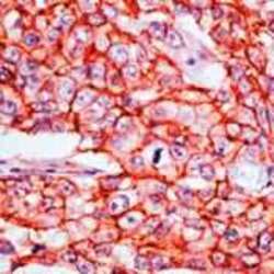
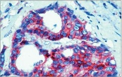
- Formalin-fixed, paraffin-embedded human breast carcinoma stained with Catenin beta antibody using peroxidase-conjugate and AEC. Note membrane staining of tumor cells.

- Submitted by
- Invitrogen Antibodies (provider)
- Main image
- Experimental details
- Immunohistochemical analysis of beta-Catenin in tumor membranes of formalin-fixed, paraffin-embedded human breast carcinoma using a beta-Catenin polyclonal antibody (Product # PA5-16762).

Supportive validation
- Submitted by
- Invitrogen Antibodies (provider)
- Main image

- Experimental details
- Figure 5. MNX1-AS1 inhibited LSCC cell viability, migration and invasion via inhibiting BCL9/beta-catenin signaling. AMC-HN-8 cells in shRNA+vector group, shMNX1-AS1+vector group and shMNX1-AS1+BCL9 groups were submitted to the following assays. (A) Western blotting technology used for the detection of BCL9 and beta-catenin expression levels ( n = 3, ** P < 0.01, compared with shRNA+vector group; ## P < 0.01, compared with shMNX1-AS1+vector group). (B) MTT assay for cell viability detection ( n = 3, ** P < 0.01, compared with shRNA+vector group). (C, D) Transwell chambers used for cell migration and invasion detection ( n = 3, ** P < 0.01, compared with shRNA+vector group; ## P < 0.01, compared with shMNX1-AS1+vector group). LSCC: laryngeal squamous cell carcinoma.

- Submitted by
- Invitrogen Antibodies (provider)
- Main image

- Experimental details
- Figure 6. Knockdown of MNX1-AS1 inhibited cell tumorigenesis in LSCC cells. (A) Tumor shapes in shRNA and shMNX1-AS1 groups. (B) Tumor volumes and weights in shRNA and shMNX1-AS1 groups. (C) The levels of miR-744-5p, MNX1-AS1 and BCL9 mRNA were detected by using qRT-PCR assay in tumor tissues from the shRNA and shMNX1-AS1 groups. (D) Immunohistochemistry technology was used to detect the protein levels of BCL9, Ki-67 and beta-catenin in tumor tissues from the shRNA and shMNX1-AS1 groups. ( ** P < 0.01, compared with shRNA group.). LSCC: laryngeal squamous cell carcinoma.
