Explore
 Validate
 Learn
Learn Western blot
Antibody data
- Antibody Data
- Antigen structure
- References [15]
- Comments [0]
- Validations
- Western blot [2]
- Immunocytochemistry [1]
- Immunohistochemistry [2]
- Flow cytometry [1]
- Chromatin Immunoprecipitation [1]
Submit
Validation data
Reference
Comment
Report error
- Product number
- AF1329 - Provider product page

- Provider
- R&D Systems
- Product name
- Human/Mouse/Rat beta-Catenin Antibody
- Antibody type
- Polyclonal
- Description
- Antigen Affinity-purified. Detects human, mouse and rat beta-Catenin in Western blots.
- Reactivity
- Human, Mouse, Rat
- Host
- Goat
- Conjugate
- Unconjugated
- Antigen sequence
P35222- Isotype
- IgG
- Vial size
- 100 ug
- Concentration
- LYOPH
- Storage
- Use a manual defrost freezer and avoid repeated freeze-thaw cycles. 12 months from date of receipt, -20 to -70 °C as supplied. 1 month, 2 to 8 °C under sterile conditions after reconstitution. 6 months, -20 to -70 °C under sterile conditions after reconstitution.
Submitted references Progranulin protects the mouse retina under hypoxic conditions via inhibition of the Toll‑like receptor‑4‑NADPH oxidase 4 signaling pathway.
Amnionless-mediated glycosylation is crucial for cell surface targeting of cubilin in renal and intestinal cells.
WNT10A mutation causes ectodermal dysplasia by impairing progenitor cell proliferation and KLF4-mediated differentiation.
Dchs1-Fat4 regulation of polarized cell behaviours during skeletal morphogenesis.
ROS-induced endothelial stress contributes to pulmonary fibrosis through pericytes and Wnt signaling.
WNT1-induced Secreted Protein-1 (WISP1), a Novel Regulator of Bone Turnover and Wnt Signaling.
Integrin β1 controls VE-cadherin localization and blood vessel stability.
Osteocytes mediate the anabolic actions of canonical Wnt/β-catenin signaling in bone.
High glucose induces podocyte injury via enhanced (pro)renin receptor-Wnt-β-catenin-snail signaling pathway.
The Wnt/β-catenin pathway attenuates experimental allergic airway disease.
Blockade of Wnt signaling inhibits angiogenesis and tumor growth in hepatocellular carcinoma.
Canonical Wnt signaling negatively regulates platelet function.
Mutational analysis of the FXNPXY motif within LDL receptor-related protein 1 (LRP1) reveals the functional importance of the tyrosine residues in cell growth regulation and signal transduction.
Dickkopf-1 is a master regulator of joint remodeling.
Requirements for proximal tubule epithelial cell detachment in response to ischemia: role of oxidative stress.
You ZP, Yu MJ, Zhang YL, Shi K
Molecular medicine reports 2019 Jan;19(1):382-390
Molecular medicine reports 2019 Jan;19(1):382-390
Amnionless-mediated glycosylation is crucial for cell surface targeting of cubilin in renal and intestinal cells.
Udagawa T, Harita Y, Miura K, Mitsui J, Ode KL, Morishita S, Urae S, Kanda S, Kajiho Y, Tsurumi H, Ueda HR, Tsuji S, Saito A, Oka A
Scientific reports 2018 Feb 5;8(1):2351
Scientific reports 2018 Feb 5;8(1):2351
WNT10A mutation causes ectodermal dysplasia by impairing progenitor cell proliferation and KLF4-mediated differentiation.
Xu M, Horrell J, Snitow M, Cui J, Gochnauer H, Syrett CM, Kallish S, Seykora JT, Liu F, Gaillard D, Katz JP, Kaestner KH, Levin B, Mansfield C, Douglas JE, Cowart BJ, Tordoff M, Liu F, Zhu X, Barlow LA, Rubin AI, McGrath JA, Morrisey EE, Chu EY, Millar SE
Nature communications 2017 Jun 7;8:15397
Nature communications 2017 Jun 7;8:15397
Dchs1-Fat4 regulation of polarized cell behaviours during skeletal morphogenesis.
Mao Y, Kuta A, Crespo-Enriquez I, Whiting D, Martin T, Mulvaney J, Irvine KD, Francis-West P
Nature communications 2016 May 5;7:11469
Nature communications 2016 May 5;7:11469
ROS-induced endothelial stress contributes to pulmonary fibrosis through pericytes and Wnt signaling.
Andersson-Sjöland A, Karlsson JC, Rydell-Törmänen K
Laboratory investigation; a journal of technical methods and pathology 2016 Feb;96(2):206-17
Laboratory investigation; a journal of technical methods and pathology 2016 Feb;96(2):206-17
WNT1-induced Secreted Protein-1 (WISP1), a Novel Regulator of Bone Turnover and Wnt Signaling.
Maeda A, Ono M, Holmbeck K, Li L, Kilts TM, Kram V, Noonan ML, Yoshioka Y, McNerny EM, Tantillo MA, Kohn DH, Lyons KM, Robey PG, Young MF
The Journal of biological chemistry 2015 May 29;290(22):14004-18
The Journal of biological chemistry 2015 May 29;290(22):14004-18
Integrin β1 controls VE-cadherin localization and blood vessel stability.
Yamamoto H, Ehling M, Kato K, Kanai K, van Lessen M, Frye M, Zeuschner D, Nakayama M, Vestweber D, Adams RH
Nature communications 2015 Mar 10;6:6429
Nature communications 2015 Mar 10;6:6429
Osteocytes mediate the anabolic actions of canonical Wnt/β-catenin signaling in bone.
Tu X, Delgado-Calle J, Condon KW, Maycas M, Zhang H, Carlesso N, Taketo MM, Burr DB, Plotkin LI, Bellido T
Proceedings of the National Academy of Sciences of the United States of America 2015 Feb 3;112(5):E478-86
Proceedings of the National Academy of Sciences of the United States of America 2015 Feb 3;112(5):E478-86
High glucose induces podocyte injury via enhanced (pro)renin receptor-Wnt-β-catenin-snail signaling pathway.
Li C, Siragy HM
PloS one 2014;9(2):e89233
PloS one 2014;9(2):e89233
The Wnt/β-catenin pathway attenuates experimental allergic airway disease.
Reuter S, Martin H, Beckert H, Bros M, Montermann E, Belz C, Heinz A, Ohngemach S, Sahin U, Stassen M, Buhl R, Eshkind L, Taube C
Journal of immunology (Baltimore, Md. : 1950) 2014 Jul 15;193(2):485-95
Journal of immunology (Baltimore, Md. : 1950) 2014 Jul 15;193(2):485-95
Blockade of Wnt signaling inhibits angiogenesis and tumor growth in hepatocellular carcinoma.
Hu J, Dong A, Fernandez-Ruiz V, Shan J, Kawa M, Martínez-Ansó E, Prieto J, Qian C
Cancer research 2009 Sep 1;69(17):6951-9
Cancer research 2009 Sep 1;69(17):6951-9
Canonical Wnt signaling negatively regulates platelet function.
Steele BM, Harper MT, Macaulay IC, Morrell CN, Perez-Tamayo A, Foy M, Habas R, Poole AW, Fitzgerald DJ, Maguire PB
Proceedings of the National Academy of Sciences of the United States of America 2009 Nov 24;106(47):19836-41
Proceedings of the National Academy of Sciences of the United States of America 2009 Nov 24;106(47):19836-41
Mutational analysis of the FXNPXY motif within LDL receptor-related protein 1 (LRP1) reveals the functional importance of the tyrosine residues in cell growth regulation and signal transduction.
Zhang H, Lee JM, Wang Y, Dong L, Ko KW, Pelletier L, Yao Z
The Biochemical journal 2008 Jan 1;409(1):53-64
The Biochemical journal 2008 Jan 1;409(1):53-64
Dickkopf-1 is a master regulator of joint remodeling.
Diarra D, Stolina M, Polzer K, Zwerina J, Ominsky MS, Dwyer D, Korb A, Smolen J, Hoffmann M, Scheinecker C, van der Heide D, Landewe R, Lacey D, Richards WG, Schett G
Nature medicine 2007 Feb;13(2):156-63
Nature medicine 2007 Feb;13(2):156-63
Requirements for proximal tubule epithelial cell detachment in response to ischemia: role of oxidative stress.
Sáenz-Morales D, Escribese MM, Stamatakis K, García-Martos M, Alegre L, Conde E, Pérez-Sala D, Mampaso F, García-Bermejo ML
Experimental cell research 2006 Nov 15;312(19):3711-27
Experimental cell research 2006 Nov 15;312(19):3711-27
No comments: Submit comment
Supportive validation
- Submitted by
- R&D Systems (provider)
- Main image
- Experimental details
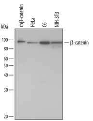
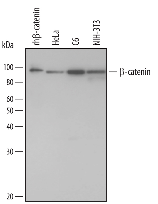
- Detection of Human/Mouse/Rat beta-Catenin by Western Blot. Western blot shows lysates of HeLa human cervical epithelial carcinoma cell line, C6 rat glioma cell line, and NIH-3T3 mouse embryonic fibroblast cell line. PVDF membrane was probed with 1 µg/mL Goat Anti-Human/Mouse/Rat beta-Catenin Antigen Affinity-purified Polyclonal Antibody (Catalog # AF1329) followed by HRP-conjugated Anti-Goat IgG Secondary Antibody (Catalog # HAF109). For additional reference, recombinant human beta-catenin (1 ng) was included. A specific band for beta-Catenin was detected at approximately 95 kDa (as indicated). This experiment was conducted under reducing conditions and using Immunoblot Buffer Group 1.
- Submitted by
- R&D Systems (provider)
- Main image

- Experimental details
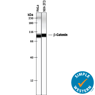
- Detection of Human and Mouse beta-Catenin by Simple WesternTM. Simple Western lane view shows lysates of HeLa human cervical epithelial carcinoma cell line and NIH-3T3 mouse embryonic fibroblast cell line, loaded at 0.2 mg/mL. A specific band was detected for beta-Catenin at approximately 94-97 kDa (as indicated) using 50 µg/mL of Goat Anti-Human/Mouse/Rat beta-Catenin Antigen Affinity-purified Polyclonal Antibody (Catalog # AF1329) followed by 1:50 dilution of HRP-conjugated Anti-Goat IgG Secondary Antibody (Catalog # HAF109). This experiment was conducted under reducing conditions and using the 12-230 kDa separation system. Non-specific interaction with the 230 kDa Simple Western standard may be seen with this antibody.
Supportive validation
- Submitted by
- R&D Systems (provider)
- Main image
- Experimental details
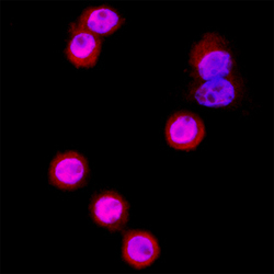
- beta-Catenin in SW480 Human Cell Line. beta-Catenin was detected in immersion fixed SW480 human colorectal adenocarcinoma cell line using Goat Anti-Human/Mouse/Rat beta-Catenin Antigen Affinity-purified Polyclonal Antibody (Catalog # AF1329) at 15 µg/mL for 3 hours at room temperature. Cells were stained using the NorthernLights™ 557-conjugated Anti-Goat IgG Secondary Antibody (red; Catalog # NL001) and counterstained with DAPI (blue). Specific staining was localized to cytoplasm and nuclei. View our protocol for Fluorescent ICC Staining of Cells on Coverslips.

Supportive validation
- Submitted by
- R&D Systems (provider)
- Main image
- Experimental details
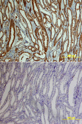
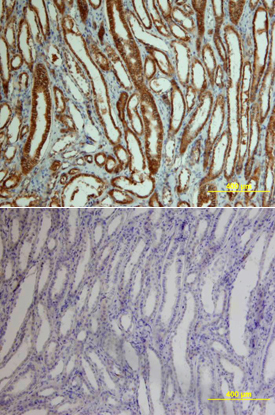
- beta-Catenin in Human Kidney Cancer Tissue. beta-Catenin was detected in immersion fixed paraffin-embedded sections of human kidney cancer tissue using Goat Anti-Human/Mouse/Rat beta-Catenin Antigen Affinity-purified Polyclonal Antibody (Catalog # AF1329) at 15 µg/mL overnight at 4 °C. Tissue was stained using the Anti-Goat HRP-DAB Cell & Tissue Staining Kit (brown; Catalog # CTS008) and counterstained with hematoxylin (blue). Lower panel shows a lack of labeling if primary antibodies are omitted and tissue is stained only with secondary antibody followed by incubation with detection reagents. View our protocol for Chromogenic IHC Staining of Paraffin-embedded Tissue Sections.
- Submitted by
- R&D Systems (provider)
- Main image

- Experimental details
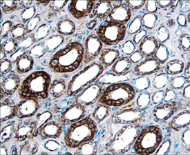
- beta-Catenin in Human Kidney Cancer Tissue. beta-Catenin was detected in immersion fixed paraffin-embedded sections of human kidney cancer tissue using 15 µg/mL Goat Anti-Human/Mouse/Rat beta-Catenin Antigen Affinity-purified Polyclonal Antibody (Catalog # AF1329) overnight at 4 °C. Tissue was stained with the Anti-Goat HRP-DAB Cell & Tissue Staining Kit (brown; Catalog # CTS008) and counterstained with hematoxylin (blue). Specific labeling was localized to epithelial cells in collecting tubules in the medulla. View our protocol for Chromogenic IHC Staining of Paraffin-embedded Tissue Sections.
Supportive validation
- Submitted by
- R&D Systems (provider)
- Main image
- Experimental details
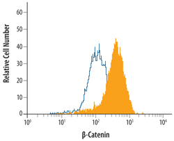
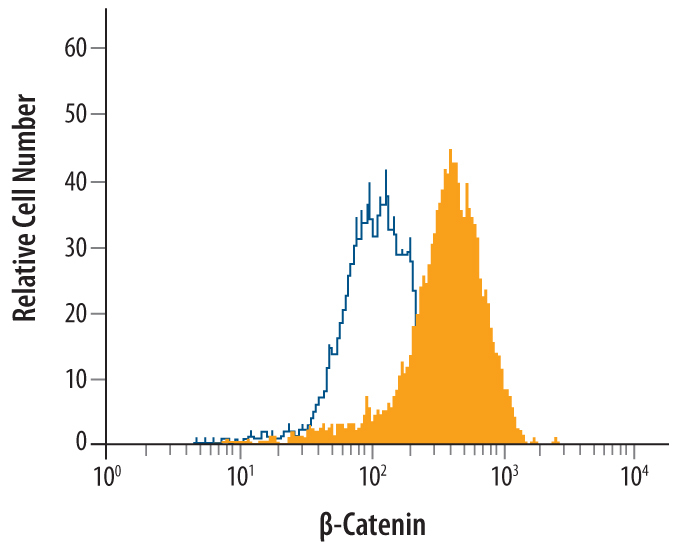
- Detection of beta-Catenin in HeLa Human Cell Line by Flow Cytometry. HeLa human cervical epithelial carcinoma cell line was stained with Goat Anti-Human/Mouse/Rat beta-Catenin Antigen Affinity-purified Polyclonal Antibody (Catalog # AF1329, filled histogram) or control antibody (Catalog # AB-108-C, open histogram), followed by Allophycocyanin-conjugated Anti-Goat IgG Secondary Antibody (Catalog # F0108). To facilitate intracellular staining, cells were fixed with paraformaldehyde and permeabilized with saponin.
Supportive validation
- Submitted by
- R&D Systems (provider)
- Main image
- Experimental details
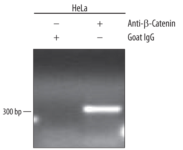
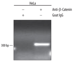
- Detection of beta-Catenin-regul-ated Genes by Chromatin Immunoprecipitation. HeLa human cervical epithelial carcinoma cell line were fixed using formaldehyde, resuspended in lysis buffer, and sonicated to shear chromatin. beta-Catenin/ DNA complexes were immuno-precipitated using 5 μg Goat Anti-Human/Mouse/Rat beta-Catenin Antigen Affinity-purified Polyclonal Antibody (Catalog # AF1329) or control antibody (Catalog # AB-108-C) for 15 minutes in an ultrasonic bath, followed by Biotinylated Anti-Goat IgG Secondary Antibody (Catalog # BAF109). Immuno-complexes were captured using 50 μL of MagCellect Streptavidin Ferrofluid (Catalog # MAG999) and DNA was purified using chelating resin solution. The SU(Z)12 promoter was detected by standard PCR.